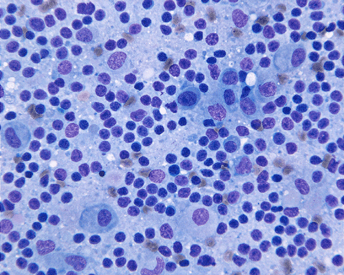

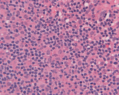
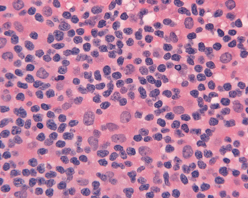
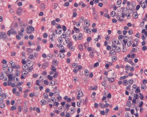
Squash
DiffQuick
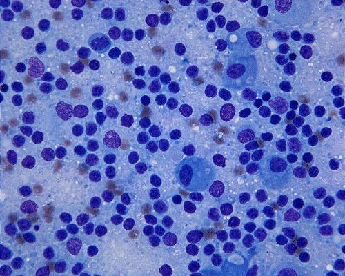
Squash
DiffQuick
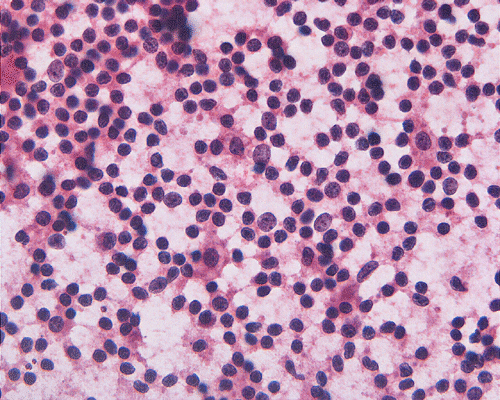
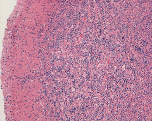
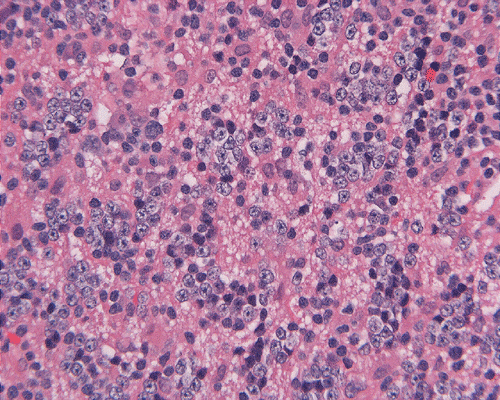
Squash
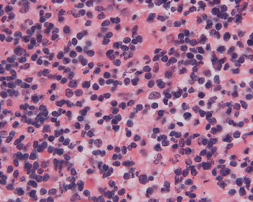
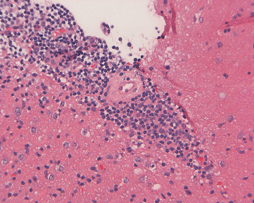
Frozen
Frozen
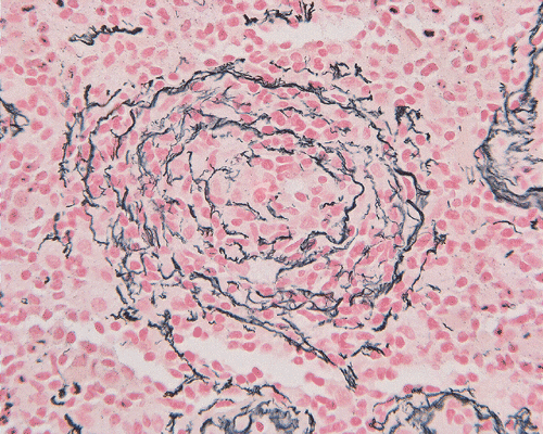
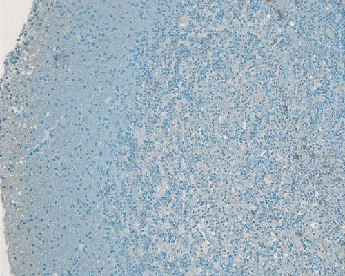
Reticuin
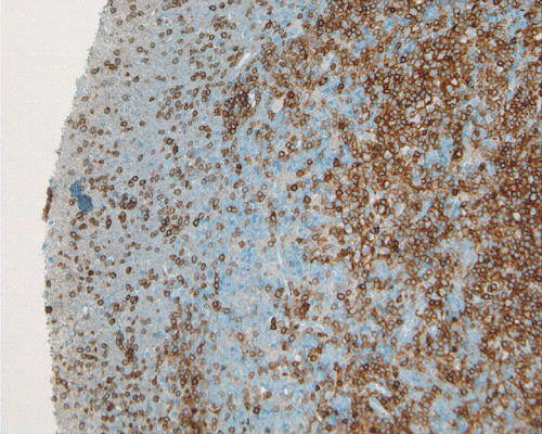
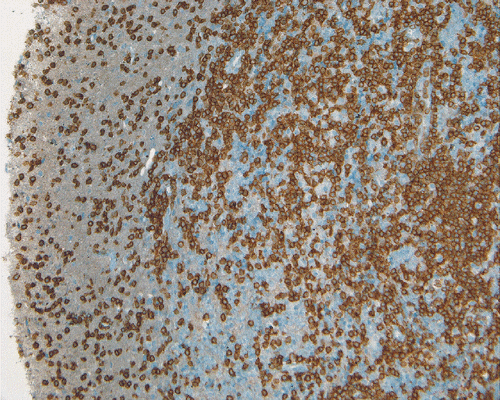
LCA
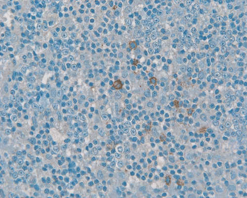
LCA
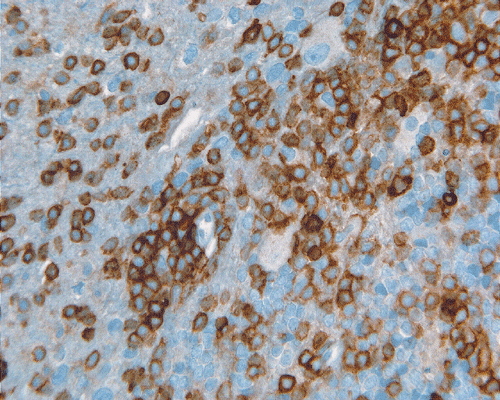
CD3
CD3
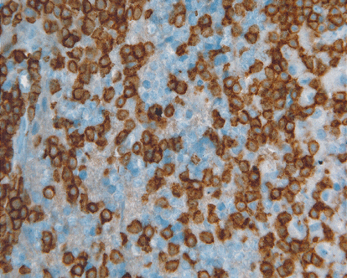
CD20
CD20
| A 75 year-old Man with
Progressive Weakness and Multiple Lesions in the Brain. May, 2006, Case 605-1. Home Page |
Brandon Guthery, M.D., Kar-Ming Fung, M.D., Ph.D. Last update: May 31, 2006.
Department of Pathology, University of Oklahoma Health Sciences Center, Oklahoma City, Oklahoma
Clinical information: The patient was a 75 year-old, right handed man who developed some progressive weakness over the last few month. There was also some right facial weakness. His weakness progressed to the point the he had multiple falling. The patient also complained of lost of balance, generalized weakness, and lost of coordination.. There was no complain of headache. There was also a history of colon cancer with surgery 6 years before the current presentation. He also had remote tonsillectomy and adenoidectomy.
On physical examination, the patient was alert to time, place, person and current events. The speech and compresension were fluent and intact. No cranial nerve abnormalities were found. Systemic workup did not reveal any other mass or space occupying lesion. On MRI, there were several intra-axial, diffusely and irregularly enhancing masses in the right cerebral hemisphere, right cerebellar peduncle, and the pons. A biopsy was performed. Representative photomicrographs are show in the followings:
|
|
|
|
|
|
|
|
A. Squash DiffQuick |
B. Squash DiffQuick |
C. Squash |
D. Frozen |
E. Frozen |
F. |
|
|
|
|
|
|
|
| G. | H. | I. | J. | K. |
L. Reticuin |
|
|
|
|
|
|
|
|
M. LCA |
N. LCA |
O. CD3 |
P. CD3 |
Q. CD20 |
R. CD20 |
Pathology of the case:
Comment: The initial clinical impression was metastases because of the history of colon cancer and multiple, irregular, enhancing lesions. The morphology and immunohistochemical profile were suggestive of a T-cell lymphoma and the diagnosis was confirmed by molecular pathology studies.
| DIAGNOSIS: Primary T-cell lymphoma of the brain. |
Discussion: Introduction Etiology Clinical Manifestation Neuroimaging Pathology Molecular Pathology
Primary central nervous system lymphoma (PCNSL) is a rare tumor. Before the advent of modern diagnostic techniques including immunohistochemistry and molecular pathology techniques, its nature was not exactly clear. In fact, they were first described by Percival Bailey as “perithelial sarcoma” because of its strong tendency to develop perivascular infiltration 1. By definition, PCNSL must originate in the central nervous system but not resulted from secondary involvement by a lymphoma in other parts of the body.
In immunocompetent individuals, PCL is an uncommon tumor that occurs in eldery age group. They account for approximately 5% of all primary brain tumors and 1-2% of non-Hodgkin’s lymphoma (NHL) 2 , 3, 4. The large majority of PCNSL are of B-cell origin; T-cell lymphomas are seen rarely and have a reported 3.6- 8.5% rate of incidence in recent large series studying PCNSL 5, 6. In the largest series of PCNSL cases diagnosed in Western countries, T-cell origin has been reported in 8 (2%) of 370 cases 7. The male to female ratio is 1.5-2:1. Primary Hodgkin’s lymphoma is almost unknown in the central nervous system. Studies have shown that the prognosis for patients with T-Cell PCNSL is similar to that for B-cell PCNSL, with cytologic type (small vs. large) having no effect on survival 10.
In immunocompromised individuals, it can affect any age group and the time of onset is related to both the time of onset and type of immunosuppression or immunodeficiency. In general, there is a peak in patients under 10 years of age which are probably resulted from congenital immunodeficiency. Another peak occurs at around the 4th decade which is probably related to HIV infection, immunosuppression due to solid organ transplantation, and other causes of immunosuppression. Theoverall survival for treated patients is much better for patients without AIDS than for those with AIDS. [See related case1] [See related case 2] [See related case 3]
Multiple theories exist on the origin of PCNSL. One proposed theory is based upon the immunologically privileged environment in the brain. This theory suggests that the malignancy develops outside the CNS originally and the immune system eliminates any peripheral involvement. An argument against the immunoprivileged theory is that patients with PCNSL do not develop lymphoma in other immunologically privileged sites, such as the testes.
Neurological symptoms present at diagnosis depend on the location of the lesion. Similar to many other space occupying lesions, the symptoms are quite non-specific. Focal motor defects are the most common symptom. Changes in personality, cognitive difficulties, and headache are common. Seizures can be seen as well. The average time from onset of symptoms to diagnosis is weeks to months, but it may be longer if personality change is the only presenting symptom 8.
The MRI findings are quite different in PCNSL occurring with and without association with AIDS, PCNSL that occurs in AIDS patients are often multifocal and centrally necrotic and appear as ring-enhancing lesions. The later features often lead to the clinical impression of a glioblastoma or metastases.
In PCNSL that are not occurring in AIDS setting, they are also common to be multiple. In particular, they tend to present as multiple, symmetrical, bilateral and subependymal foci. In contrast to those occurring in AIDS, these tumor are typically non-necrotic and without ring enhancement. In fact, enhancement is rather homogeneous.
Primary CNS lymphomas (PCNSL) can be firm, friable, granular, centrally necrotic, focally hemorrhagic, grey-tan, yellow, or virtually indistinguishable from the adjacent neuropil. Some tumors are well-delineated, like a metastasis; while some present as diffusely infiltrating forms without evidence of mass lesions. Multiple lesions are seen in about one-third of the patients. Typically, they are deep-seated in the cerebral hemispheres and are adjacent to the ventricules. Less commonly PCNSL are superficially located and one study showed an increased incidence of this with T-cell PCNSL 9. A small proportion of them are limited only to the dura.
Reference
Jellinger K, Seitelberger F . Malignant lymphomas of the nervous system. International Symposium Wien, 1974 Acta Neuropathol Suppl 1975 6: 1-301.
Behin A, Hoang-Xuan K, Carpentier AF, Delattre JY. Primary brain tumours in adults. Lancet. 2003 361:323-31.
DeAngelis LM, Currie VE, Kim JH, Krol G, O'Hehir MA, Farag FM, Young CW, Posner JB. The combined use of radiation therapy and lonidamine in the treatment of brain metastases. J Neurooncol. 1989 7:241-7.
Miller DC, Hochberg FH, Harris NL, Gruber ML, Louis DN, Cohen H. Pathology with clinical correlations of primary central nervous system non-Hodgkin's lymphoma. The Massachusetts General Hospital experience 1958-1989. Cancer. 1994 74:1383-97.
Bataille B, Delwail V, Menet E, Vandermarcq P, Ingrand P, Wager M, Guy G, Lapierre F. Primary intracerebral malignant lymphoma: report of 248 cases. J Neurosurg. 2000 92:261-6.
Hayabuchi N, Shibamoto Y, Onizuka Y. Primary central nervous system lymphoma in Japan: a nationwide survey. Int J Radiat Oncol Biol Phys. 1999 44:265-72.
Ferreri AJ, Reni M, Pasini F, Calderoni A, Tirelli U, Pivnik A, Aondio GM, Ferrarese F, Gomez H, Ponzoni M, Borisch B, Berger F, Chassagne C, Iuzzolino P, Carbone A, Weis J, Pedrinis E, Motta T, Jouvet A, Barbui T, Cavalli F, Blay JY. A multicenter study of treatment of primary CNS lymphoma. Neurology 58:1513-1520, 2002.
Herrlinger U, Schabet M, Bitzer M, Petersen D, Krauseneck P. Primary central nervous system lymphoma: from clinical presentation to diagnosis. J Neurooncol. 1999 43:219-26.
Choi JS, Nam DH, Ko YH, Seo JW, Choi YL, Suh YL, Ree HJ. Primary central nervous system lymphoma in Korea: comparison of B- and T-cell lymphomas. Am J Surg Pathol. 2003 27:919-28.
Shenkier TN, Blay JY, O'Neill BP, Poortmans P, Thiel E, Jahnke K, Abrey LE, Neuwelt E, Tsang R, Batchelor T, Harris N, Ferreri AJ, Ponzoni M, O'Brien P, Rubenstein J, Connors JM. Primary CNS lymphoma of T-cell origin: a descriptive analysis from the international primary CNS lymphoma collaborative group. J Clin Oncol. 2005 23:2233-9.