EMA
GFAP
| A 10 year-old Girl with a
Cerebral Hemispheric Mass and Hydrocephalus. June, 2006, Case 606-1. Home Page |
Eric Harp, D.O., Kar-Ming Fung, M.D., Ph.D.
Department of Pathology, University of Oklahoma Health Sciences Center, Oklahoma City, OK, Last update May 30, 2006
Clinical information: This patient was a 10 year-old girl with unremarkable birth history and normal development. Her academic performance declined substantially the year before presentation and she also complained of headache. An MRI scan reviewed a large cerebral hemispheric, parenchymal mass that bulged into the ventricle and compressed the foramen of Monro. She also had hydrocephalus that was resulted from the mass. Surgery was performed to remove the mass and yielded the following representative pictures:
|
|
|
|
|
|
|
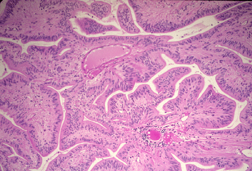
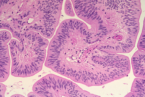
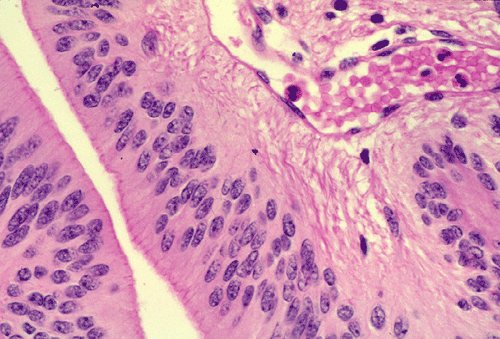
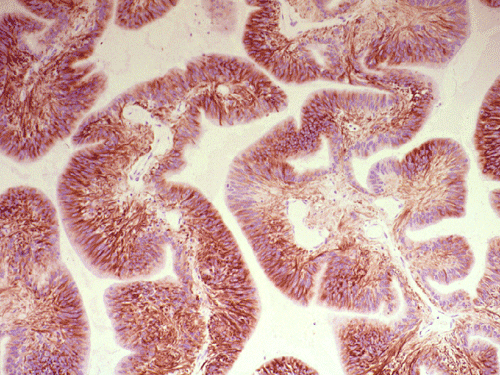
| A. | B. | C. |
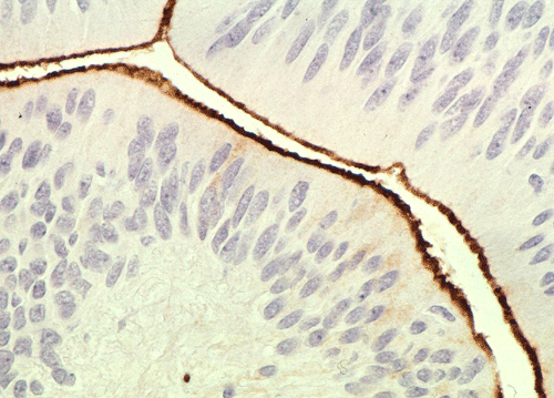
D. EMA |
E. GFAP |
Pathology of the case:
The histopathology of the tumor is fairly similar in different parts of the of the tumor and the papillary structures are the obvious (Panel A). On medium and high magnification (Panel B and C respectively), the cores of the papillary structure is composed of thin walled blood vessels. The tumor cells appear to appear in a pattern reminiscent of the pseudocolumnar arrangement of the respiratory respiratory epithelium except that no basement membrane can be identified. The cores are also not composed of collagenous tissue but by fibrillary process that appear to extends from the nuclei. A zone of hyponuclear area is present between the nuclei and the vascular channels (delimited by arrows in Panel C). No cilia are noted on the surface epithelium and no mucin production is noted. The cytoplasmic border is very indistinct. The nuclei appear to be rather bland appearing and mitotic figures are not readily seen. There is neither endothelial proliferation nor necrosis.
On immunohistochemistry, the luminal boder of the cells are strongly positive for epithelial membrane antigen (EMA) (Panel D). The tumor cells are also strongly positive for glial fibrillary acidic protien (GFAP) (Panel E).
| DIAGNOSIS: Papillary Ependymoma (WHO grade II). |
Discussion: General Information Genetics Pathology Differential Diagnosis
General Information:
In the World Health Organization (WHO) classification 1, ependymomal tumors are classified as follows:
Supependymoma (WHO grade I) [Click here to see a case]
Myxopapillary ependymoma (WHO grade I) [Click here to see a case]
Ependymoma (WHO grade II)
variants: cellular ependymoma, clear cell ependymoma, tanycytic ependymoma, papillary ependymoma
Anaplastic ependymoma (WHO grade III)
Ependymoma (WHO grade II) is a rather common tumor that affects both sexes equally. They are generally seen in the first two decades of life but can occur at any age. Ependymomas comprise about 10% of pediatric central nervous system (CNS) neoplasms and 5% of all gliomas regardless of age group. About 30% of them are seen in children under 3 years of age. In fact, medulloblastoma, astrocytoma particularly pilocytic astrocytomas, choroid plexus tumors and ependymomas are the four most commonly seen tumors of the CNS in children and infants. Ependymoma is also the most common spinal cord neuroepithelial lesions, accounting for 50-60% of spinal cord gliomas.
Ependymomas occur in decreasing frequency in these sites: fourth ventricle, lateral ventricle, third ventricle, aqueduct, spinal cord, cauda equina, extraventricular hemisphere. Most of the sites are associated with the ventricular system and, therefore, hydrocephalus is not an uncommon finding.While spinal ependymomas are usually found in adults, cerebral ependymomas are usually found in children.
Ependymomas have a strong tendency to recur at the primary site and gross total resection of the primary tumor remains an important independent positive prognostic factor. They also have a tendency to disseminate through the cerebral spinal fluid and, unfortunately, this sinister behavior cannot be always predicted with their histologic features or histologic grade. Prognosis in children is usually worse than that in adults. Incomplete resection, leptomeningeal dissemination, and patients age under 3 year are both unfavorable prognostic factors. The 5-, 10-, and 15-year overall survival rates for ependymoma is 71.4%, 63.5%, and 63.5% in patients with craniospinal radiotherapy respectively 2.
The prognosis is also related to the location of the tumor. In general, spinal ependymomas have better prognosis than intrcranial ependymomas. Tumors in the posterior fossa and 4th ventricle is often associated with hydrocephalus, compression of the brainstem and spinal cord. They also have an increased tendency to disseminate along the spinal cord. In addition, hemorrhage is not uncommon and would lead to rapid expansion of the tumor, compression of the brainstem and sudden death.
Patients with neurofibromatosis 2 (NF2) have increased incidence of ependymomas, sometimes with multiple intramedullary ependymomas, and other proliferative ependymal lesions. Mutations of NF2 gene and loss of chromosome 22q occur preferentially in intramedullary spinal ependymomas 3. A tumor suppressor gene may be present in chromosome 22pter-22q11.2, other gene involved may be in chrormosome 6q and X chromosome. There is, however, no consistent genetics abnormalities that are associated with ependymoma.
Grossly, ependymomas are generally well demarcated intraparenchymal tumors. Some of the cases may protrude into the ventricles to give a cauliflower-like pattern. They are usually soft with varying red to brown to gray color. A papillary lesion would show excresences. Necrosis is not a feature of low grade tumor but can be seen in high grade tumor. Hemorrhage is not uncommon. Ependymomas arising in the posterior fossa or 4th ventricles have a particular tendency to exrtend through foramen of Luschka, Magendie, and then downward through foramen magnum, resulting in caudal tongue-like projections of tumor which compress dorsal and lateral aspects of the upper cervical cord (so called "plastic ependymoma").
Microscopically, ependymoma is a well know imposter. They are usually of moderate cellularity. However, the cellularity can be high enough to suggest a medulloblastoma or low enough to suggest a pilocytic astrocytoma, particularly in frozen sections.
The diagnostic pattern of ependymoma is best visualized with very low magnification under the microscope [Click here to see pictures of classic ependymomas]. Ependymomas are characterized by a perivascualr radial formation of isomorphic tumor cells with a nuclear-free zone that form a cuff around the vessels is an important diagnostic feature. The tumor cells have fibrillary cytoplasmic processes that taper into this cuff. Ependymal canals are tubular structures lined by ependymal cells. A basement membrane is characteristic missing and the cells with distinct epithelial morphology merges distinctly with the underlying tumor cells. Although they are not common features but is of great diagnostic value when being found. Mitotic figures, if present, should only be sporadically seen. Cystic component and calcifications may be present. Significant pleomorphism, increased mitotic activities, and necrosis are suggestive of anaplastic ependymoma (WHO grade III).
Clear cell ependymoma refer to a variant that have clear cytoplasm. These tumors must not be confused with oligodendrogliomas and central neurocytomas. Cellular ependymoma refer to a variant that have increased cellularity. Tanycytic ependymoma is a rare fibrillar variant of ependymoma characterized by streams of piloid or hair-like, cells having "ependymal" nuclei. While some of them main the appearance of classic ependymoma with perivascular cuffing, others may look like an “intra-axial schwannoma”.
Papillary ependymoma, as illustrated in this case, is a rare variant. These tumors typically appear as papillary fronds covered by multilayered ependymal tumor cells. The most superficial layer may have epithelium-like surfaces similar to those of ependymal rosettes. These tumors must be distinguished from metastatic papillary carcinomas and choroids plexus tumors. One salient feature is that papillary ependymomas have no basement membrane. The superficial epithelial tumor cells merge imperceptibly with the underneath tumor cells. Papillary ependymoma can express cytokeratin. It is not rare to see focal papillary formation in an otherwise classic ependymoma. However, tumors composed predominantly of papillary structure as illustrated in this case is uncommon.
On immunohistochemistry, ependymomas usually demonstrate widespread positivity for glial fibrillary acidic protein (GFAP). The tumor cells, particularly the luminal surface of ependymal canals or papillary ependymoma are positive for epithelial membrane antigen. Over 80% of ependymomas are positive for CD99 4, 5. However, a small number of other neuroepithelial tumors are also positivie for CD99, this feature is not a full proof diagnostic help. For low-grade ependymomas the progression free survival time is significantly shorter with Ki-S1 labeling index over 5%, and for tenascin, VEGF, and EGFR positivity on immunohistochemistry. For high-grade ependymomas PFS was found to be significantly reduced for age under 16 years, subtotal tumor removal, p27 labeling index under 20%, p53 positivity, and for apoptotic index under 1% 6. Decreased expression of p14ARF protein expression is also related to aggressive biological behavior 7.
At ultrastructural level, the presence of membrane junctional complex, microrosette, and 9+2 cilia are diagnostic features for ependymomas. [See a related question]
Differential Diagnosis:
The pathologic features in this case clearly point to a papillary tumor arising in the cerebral hemisphere. The first issue of concern is whether this is a primary or secondary tumor. Metastatic papillary carcinoma in a 10 year-old girl is very unusual, particularly without a confirmed history of papillary carcinoma in locations outside the brain. For all practical purpose, this tumor should be viewed as a primary tumor.
The major differential diagnosis is choroid plexus papilloma. Most choroid plexus papillomas occur as an exophytic mass that usually protrudes into the ventricle. The imaging results of a hemispheric mass that bulged into the ventricles is not the most common picture of a choroid plexus papilloma. In addition, the degree of pleomorphism and the age of this patient are not compatible with choroid plexus carcinoma; most of them are seen in infants. The fact that the tumor cells have a palisading arrangement, a nuclear free mantle zone around blood vessels, and the absence of basement membrane suggest a diagnosis of papillary ependymoma. Most importantly, there is a lack of basement membrane to support the diagnosis of a choroid plexus papilloma. Immunohistochemistry, choroid plexus papilloma are often positive for transthyretin and synaptophysin and these makers are negative in papillary ependymomas [Click here to see a table for comparison].
Reference:
Kleihues P. Cavenee W. WHO Classification of Tumours: Tumours of the Nervous System. IARC press. 2000.
Paulino AC, Wen BC, Buatti JM, Hussey DH, Zhen WK, Mayr NA, Menezes AH. Intracranial ependymomas: an analysis of prognostic factors and patterns of failure. Am J Clin Oncol. 2002 Apr;25(2):117-22.
Ebert C, von Haken M, Meyer-Puttlitz B, Wiestler OD, Reifenberger G, Pietsch T, von Deimling A. Molecular genetic analysis of ependymal tumors. NF2 mutations and chromosome 22q loss occur preferentially in intramedullary spinal ependymomas. Am J Pathol. 1999 Aug;155(2):627-32.
Choi YL, Chi JG, Suh YL. CD99 immunoreactivity in ependymoma. Appl Immunohistochem Mol Morphol. 2001 Jun;9(2):125-9.
Kawano N, Yasui Y, Utsuki S, Oka H, Fujii K, Yamashina S. Light microscopic demonstration of the microlumen of ependymoma: a study of the usefulness of antigen retrieval for epithelial membrane antigen (EMA) immunostaining. Brain Tumor Pathol. 2004;21(1):17-21. Erratum in: Brain Tumor Pathol. 2004;21(2):102-3.
Korshunov A, Golanov A, Timirgaz V. p14ARF protein (FL-132) immunoreactivity in intracranial ependymomas and its prognostic significance: an analysis of 103 cases. Acta Neuropathol (Berl). 2001 Sep;102(3):271-7.
Korshunov A, Golanov A, Timirgaz V. p14ARF protein (FL-132) immunoreactivity in intracranial ependymomas and its prognostic significance: an analysis of 103 cases. Acta Neuropathol (Berl). 2001 Sep;102(3):271-7.