| A 28 year-old Man with a Small
Nodule on his Tongue. August, 2006, Case 608-1. Home Page |
Kar-Ming Fung, M.D., Ph.D. Last update August 30, 2006.
Department of Pathology, University of Oklahoma Health Sciences Center, Oklahoma City, OK
Clinical information: The patient was a 28 year-old man with a small 0.5 cm mucosa covered nodule on the side of his tongue. The polyp was excised for pathologic examination. The followings are the representative images:
|
|
|
|
|
|
|
|
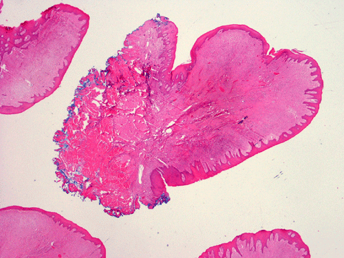
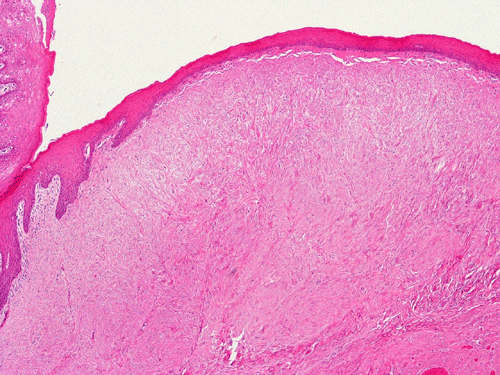
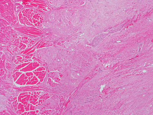
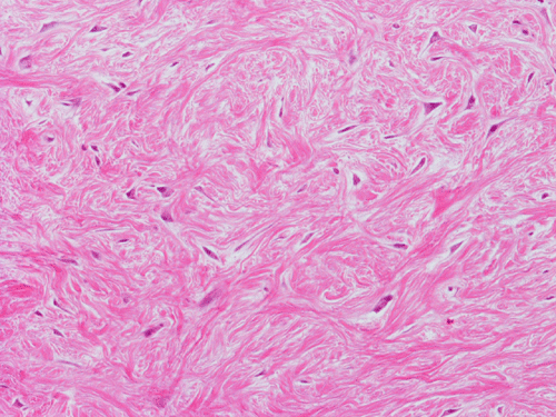
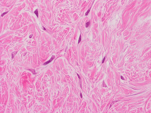
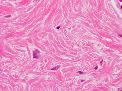
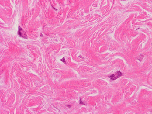
| A. | B. | C. | D. | E. | F. | G. |
Pathology of the case:
Grossly, the specimen, 1.2 cm in greatest dimension, consists of a nodule of fibrous tissue with tan brown muscle attached to one side. It was submitted entirely for microscopic examination after serial sectioning.
On low magnification, the lesion is a mucosa covered polypoid nodule that is free of ulceration, erosion, and inflammation (Panel A). The resection margin contains muscle of the tongue (M in Panel A). There is some flattening of the epithelial ridges overlying the nodule (Panel B). The nodule does not invade into the skeletal muscle of the tongue (Panel C). On medium magnification, the nodule is composed predominantly of fibrous tissue that contains a low density of spinlde shaped, fibroblast like cells (Panel D and E). On high magnification, many of these spindle cells appear triangular in shape and contains two to three nuclei but are not associated with atypia or nuclear pleomorphism (Panel F, G). Mitoses and necrosis are not readily seen. No hypercellular region is present in the entire specimen.
| DIAGNOSIS: Giant cell fibroma. |
Discussion:
General Information:
This family of lesion is also known as "bite" or "irritation"fibroma, fibroepithelial or fibrovascular polyp. They are mostly seen in the buccal mucsal and tongue as a result of trauma by biting or in the gingiva as a result of plaque accumulation or irritation by a denture. Grossly, they typically appear as a nodule or a polypoid lesion.
Pathology:
This entity is essentially a nodular proliferation of fibrous tissue with scar like pathologic changes. A variable amount of vascularity is resent. Reactive changes in the overlyig epidermis including ulceration and granulation can occur. The fibrous stroma can be scleortic to myxoid. Entrapped muscle fiber can be present. Entrapped cartilage suggestive of chondroid metaplasia can be seen.
Histologically, giant cell
fibroma is a fibroma decorated by scattered triangular fibroblast like cells
containing a few nuclei. Sclerotic fibromas are suggestive of tuberous sclerosis
which is associated with heterozygosity of PTEN gene.
Sclerotic fibroma (also called storiform collagenoma)
is charactered by a dense, hyalinized bands of thick collagen fibers resembling
keloid with fusiform fibroblasts in a slighly storiform pattern. They can be
positive for factor XIIIa and CD34.
Reference: