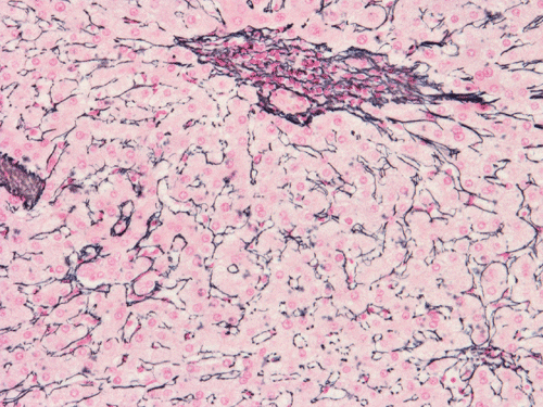
Reticulin
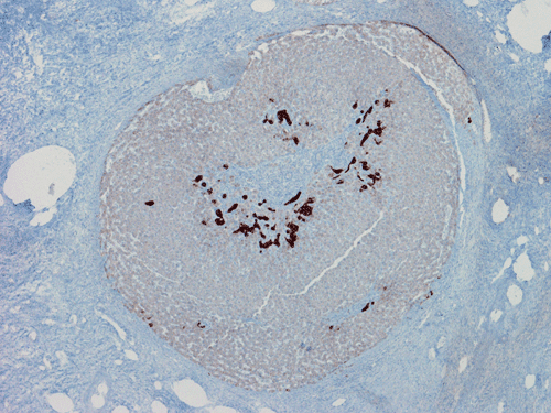
CK7
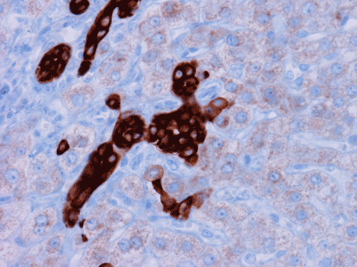
CK7
| A 21 year-old Woman with a
Hepatic Mass. October, 2006, Case 610-1. Home Page |
Jinous Saremian, M.D., Cheng Z. Liu, M.D. Ph.D. Last update: October 31, 2008.
Department of Pathology, University of Oklahoma Health Sciences Center, Oklahoma City, Oklahoma
Clinical information: The patient was a 21 year-old female who complained of multiple episodes of epigastric discomfort with nausea and vomiting. An ultrasound examination was performed and revealed a hepatic mass in the left lobe of liver. A CT scan revealed a well defined mass that occupied most of the left lobe of the liver. The mass was excised. The followings are representative images of the specimen.
|
|
|
|
|
|
|
| A. | B. | C. | D. | E. | F. |
|
|
|
|
|
|
|
| G. | H. |
I. Reticulin |
J. CK7 |
K. CK7 |
Pathology of the case:
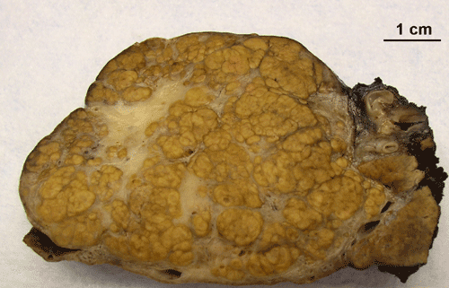
The gross specimen as illustrated in Panel A is 242 gram and 11.0 x 8.0 x 5.5 cm. The lesion is a well defined nodule that can be easily shelled out during surgery. The cross section is composed of pale yellow-brown nodules of similar size and separated by thin fibrous septa. A large a stellate fibrotic scar (C in Panel A) is present. Also note that the tumor is well demarcated from the surrounding liver parenchyma (arrow in Panel A)
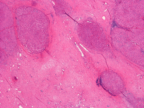
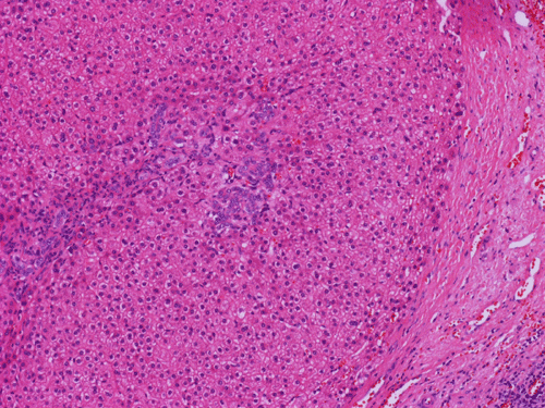
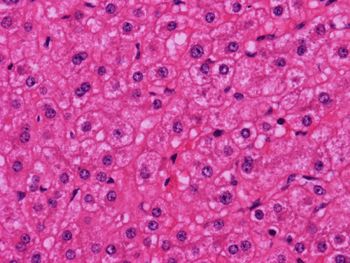
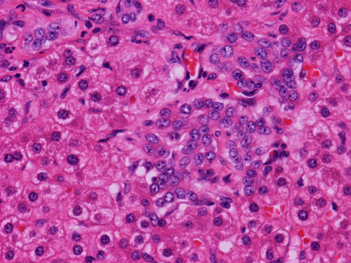
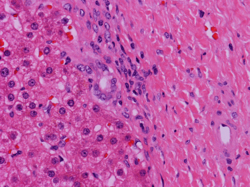
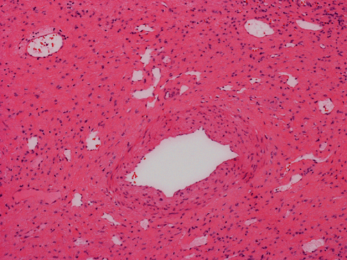
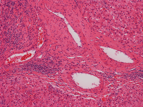
Histologically, the grossly notable nodules correspond to islands of cells separated by dense fibrous tissue (Panel B). On medium magnification, the islands of cells have smooth, pushing margins (Panel C). The cells are quite similar to normal hepatocytes except that these islands do not have the general architecture of normal liver (Panel D). The liver plate varies from one to three cell thick is well demonstrated by reticulin stain (Panel I). In some areas there are some entrapped ductular structure consistent with bile ductules (Panel E and F) and they are well demonstrated by immunohistochemistry for cytokeratin 7 (Panel J and K). Most of these ductules are found around the fibrous scar or septa or at the edge of the nodules. Many abnormally large blood vessels are also noted and many of them are seen in the fibrous central scar (Panel G and H).
| DIAGNOSIS: Focal nodular hyperplasia. |
Discussion:
General Information
Focal nodular hyperplasia (FNH) is the second most common benign hepatic mass after hemangioma. FNHs are found mainly in young women in their third and forth decades 1, 2. Around 5-15% of cases occur in males and 7-15% in pediatric age group. Increased in incidence with the use of oral contraceptives is well known. It has a well-known association with the use of oral contraceptives. Unlike adenomas, FNH is thought to be developed as a response to increased blood flow in the abnormal vessels in FNH but there is a speculation that the use of contraceptives may increase their size. FNHs are often discovered as incidental findings during surgery, unrelated radiographic or ultrasonic examination of the liver, and during autopsies 1, 2. Symptomatic FNHs may manifest with abdominal fullness, pain, discomfort, anorexia or fatigue 3. The presence of the central scar is a great help in diagnosis by MRI and ultrasound. FNH has no known malignant potential.
FNH is usually solitary but about 20-30% of the cases are multifocal. Multiple FNH syndrome refers to a condition that at least two FNHs are present and is associated with one or more other lesions which include glioma, meningioma, hepatic hemangioma and vascular malformations. FNH is also associated with hepatic cavernous hemangioma, glycogen storage disease type Ia, or portal hypertension. By definition, the hepatic parenchyma surrounding FNH should be normal, therefore hepatic nodules that are found in patients with Osler-Weber-Rendu disease, Budd-Chiari syndrome, and cirrhosis (MRNs) are not FNH although these nodules closely mimic FNH clinically.
Pathology:
Grossly, FNHs have a rather characteristic look. They are light brown, well defined, unencapsulated, lobulated, subcapsular nodules up to a few centimeter in greatest dimension. About two-third of FNHs occur in the right lobe and they are usually under 5 cm in greatest dimension.The cut surface is solid and reminiscent of normal liver. A signature central, stellate, fibrous scar that radiates out into the mass and divides the mass in to small nodules reminiscent of cirrhosis is typically present. The size of the scar is usually large enough to be obvious. The diagnosis of FNH should be questioned if this scar is missing. A detailed and mindful examination of the specimen should be able to find smaller scars in many questionable cases. In FNHs smaller than 1 cm, the central scar may not be easily detectable 4. Neither necrosis nor hemorrhage is typical for FNHs.
The histopathologic triad of FNH is bland hepatocytes without abnormal architecture, preserved reticulin network, the presence of bile ductules, central scar with fine fibrous septa radiating out, and the presence of large and abnormal blood vessels surrounded by a zone of connective tissue.
The central scar is composed of dense collagenous fibrous tissue with a variable number of tortuous thick-walled arteries. Fibromascular hyperplasia and myxoid degeneration are often found in these vessels.The areas away from the scaring portion and the fibrotic septa would look like normal liver at scanning magnifications. The key feature at low magnification is that no normal portal triads, no central vein or portal arteries are present. Instead, bile ductules are usually evident and often associated with chronic inflammatory cell infiltration.
Histologically, the cells in FNH are almost identical to the normal hepatocytes surrounding hepatocytes. The reticulin network is preserved and the liver plate varies from one to three cells thick. The cells in FNA may possess increased cytoplasmic glycogen content, focal steatosis, bile stasis, lipofuscin, iron pigment, copper deposits and Mallory bodies 4. The central scar is composed of dense collagenous fibrous tissue with a variable number of tortuous thick-walled arteries. Fibromascular hyperplasia and myxoid degeneration are often found in these vessels.
FNH is essentially a morphologic diagnosis. Immunohistochemically, they express hepatocellular markers such as Her Par 1. They are also positive for CAM5.2, polyclonal CEA, and alpha-1- antitrypsin. CD34 is positive in endothelial cells lining the cell plates. AFP and P53 are negative. CK7 is negative in the hepatocytes but they are useful in demonstrating the proliferating bile ductules. This use, is more of educational than diagnostic purposes.
Pathogenesis:
The central scar with abnormal blood vessels is proposed to be the principal etiology of FNH and these lesions are considered hyperplastic changes in response to increased blood flow in these malformed blood vessels 5, 6, 7. Aberrant expression of angioprotein genes, involved in the regulation of vasculogenesis, may play a role in the formation of these hyperplastic and dystrophic vessels 9. The proliferating hepatocytes in most FNHs are polyclonal 8. No specific molecular markers have been identified.
Differential Diagnosis:
The major differential diagnosis include bile duct adenoma, well differentiated hepatocellular carcinoma particularly the fibrolamellar type and regenerating nodules associated with other conditions such as cirrhosis and Budd-Chiari syndrome.
FNH and adenoma share the common features of occurring in young woman during their reproductive age and are related with the use of contraceptives. Both of them share the similar cytologic features. The cells are very similar to the normal surrounding hepatocytes. The cell plate are one to three cells thick and is best demonstrated by reticulin stain. Adenoma, however, has even stronger inclination to occur in woman. FNH and adenoma, however, have different imaging and scintigraphic characteristics. FNH reveals a homogenous enhancement with a central scar on CT and MRI and normal or increased uptake on Tc-sulfur-colloid scintigraphy. While adenoma appears as a hyperintence heterogenous mass in image and has no uptake in scintigraphy. The central scar is also helpful on histologic examination. It should be noted that the scar in small FNH may not be obvious. A high level of suspicion and careful examination is mandatory. Histologically FNH usually have ductule proliferation and hyperplastic and abnormal large vessels. The ductules are usually found in the fibrovascular areas and often at the edge of the tumor. Although large vessels are often seen in adenoma, there is no significant connective tissue zone around them. In contrast, vessels in FNH are abnormally large and are surrounded by a zone of connective tissue. Adenoma can have occasional fibrous septa which tends to be discontinuous. The fibrous scar and septa in FNH is more prominent.
Both FNH and fibrolamellar hepatocellular carcinoma (FLHCC) have fibros component which can cause diagnostic confusion. In addition, both have a tendency to occur more frequently in young women. However, FL-HCC is grossly more irregular in shape than FNH. For small biopsies, correlation with MRI or ultrasound findings would be helpful. Second, the fibrosis in FLHCC does not typically appear as central radiating scar but rather heterogeneous and diffuse. The neoplastic cells may occur as irregular islands among the fibrous component. Rarely, a central scar similar to that of FNH may be present in FLHCC. Third, there is usually significant nuclear atypia in FLHCC. Their nucleoli are prominent. Fourth, although FLHCC is more circumscribed than the conventional hepatocellular carcinoma, infiltration can still be found at the periphery of FLHCC but not FNH. Fifth, liver plate architecture is not preserved in FLHCC but the liver plate in FNH is limited one to three cells thick. In addition, there may be acini formation by the tumor cells, presence of bile in tumor cells, and multinucleated tumor cells in FLHCC. Glandular formation and mucin secretion can occur in FLHCC.
Regenerative nodules associated with cirrhosis and Budd-Chiari do not have fibrous septa and are surrounded by abnormal liver with condensation of reticulin network.References:
Saul SH. Masses of the liver. In: Sternberg SS, ed. Diagnostic Surgical Pathology. 4th ed. New York, NY: Raven; 2004 1705-1774.
Lizardi-Cervera J, Cuéllar-Gamboa L, Motola-Kuba D. Focal nodular hyperplasia and hepatic adenoma: a review. Ann Hepatol. 2006 5:206-11.
Pan Y, Wang ZM, Mou LJ, Teng XD, Zheng ZJ, Ying LX. Focal nodular hyperplasia of the liver: pathological analysis of 11 cases. Hepatobiliary Pancreat Dis Int. 2004 3:199-203.
Walness I, Callea F, Craig J et al. Terminology of nodular hepatocellular lesions. International Working Party. Hepatology 1995 22:983-93.
Wanless IR, Albrecht S, Bilbao J, Frei JV, Heathcote EJ, Roberts EA. Multiple focal nodular hyperplasia of the liver associated with vascular malformations of various organs and neoplasia of the brain: a new syndrome. Mod Pathol. 1989 2:456-62.
Wanless IR, Mawdsley C, Adams R. On the pathogenesis of focal nodular hyperplasia of the liver. Hepatology. 1985 5:1194-200.
Wanless IR. Micronodular transformation (nodular regenerative hyperplasia) of the liver: a report of 64 cases among 2,500 autopsies and a new classification of benign hepatocellular nodules. Hepatology 1990 11:787-97.
Paradis V, Laurent A, Flejou JF, Vidaud M, Bedossa P. Evidence for the polyclonal nature of focal nodular hyperplasia of the liver by the study of X-chromosome inactivation. Hepatology. 1997 26:891-5.
Paradis V, Bièche I, Dargère D, Laurendeau I, Nectoux J, Degott C, Belghiti J, Vidaud M, Bedossa P. A quantitative gene expression study suggests a role for angiopoietins in focal nodular hyperplasia. Gastroenterology. 2003 124:651-9.