| A 45 year-old Man with an
Ulcer on his Hard Palate. January, 2007, Case 701-1. Home Page |
Ashish Bains, M.D., Glen D. Houston, D.D.S.
Department of Pathology, University of Oklahoma Health Sciences Center, Oklahoma City, OK Last update April 30, 2007
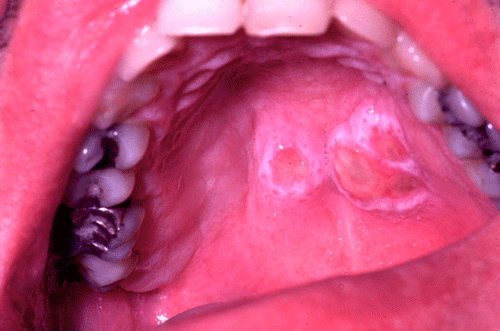
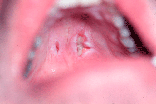
Clinical information: The patient was a 45 year-old smoker who presented to his dentist with a painless ulcer in his hard palate as illustrated below. A biopsy was performed. Representative histopathologic images are illustrated below:
|
|
|
|
|
|
|
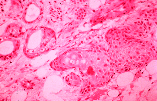
| 1. | A. | B. | C. | D. |
Pathology of the case:
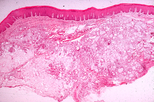
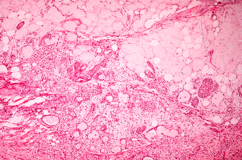
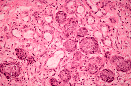
An ulcer is present in the hard palate as illustrated in (Panel 1). In the areas that are slightly taken away from the ulcer, minor salivary glands are present. The more superficially located glands seem to have lost mucin production in this scanning magnification image (black arrow in Panel A). There is also fibrous thickening of the subepidermal areas (which arrow in Panel A). On higher magnification, the mucus secreting cells are replaced by squamous epithelium (Panel B and C). Note that these squamous cell nests are well demarcated, non-infiltrative, and about the size off the mucus secreting gland. There is not significant pleomorphism in these nests and are most consistent with squamous metaplasia (Panel D).
| DIAGNOSIS: Necrotizing sialometaplasia. |
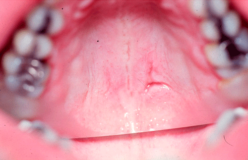
Post treatment:
The patient was treated and her ulcer healed in a few weeks time (Panel 2 and 3).
 |
 |
||||
| 2. | 3. |
Discussion:
General Information
As reflected by the name, necrotizing sialometaplasia (NS) is a lesion characterized by necrosis and metaplastic change of minor salivary gland. It is a benign, self limiting inflammatory condition that affects almost exclusively minor salivary gland with the palate as the most commonly involved site. NS is believed to be produced by pathophysiological changes following vascular ischemic insults of the salivary gland. NS was first described in 1973 by Abrams et al 1, and since then many cases have been reported in oral cavity, paranasal sinuses and other areas with minor salivary glands. The importance of this entity lies in the fact that its clinical and histological features closely mimic malignancy, particularly squamous cell carcinoma.
Clinically, most cases involve minor salivary glands with over 75% occurring in the posterior palate, of these about two-thirds are unilateral 2. The initial lesion is non-ulcerated swelling, which may or may not be associated with pain and paresthesia. It evolves into a necrotic lesion which sloughs off leaving an ulcerated base. The ulcerated lesion ranges from less than 1 cm to more than 5 cm in diameter 2. Erosion of the palatal bone may occur, but is uncommon 3. The mean age of patients with NS is 46 years and males outnumbered females by a ratio of 2 to 1 3. Cases of NS have also been reported in pediatric population 4 but they are uncommon. Unfortunately, NS can be easily misdiagnosed as mucoepidermoid carcinoma or squamous cell carcinoma, which lead to devastating surgical interventions. Correctly diagnosing NS is thus of paramount importance.
Pathology
The cause of NS, although uncertain 2, 5, is widely believed to be secondary to ischemia of the salivary glands 5, 6, 7. Many cases arise spontaneously, but history of trauma, dental injections, ill-fitted dentures, radiotherapy and previous surgery have all been described as predisposing factors. Clinically, it can mimic many malignancy. Incisional biopsy plays a critical role in making the correct diagnosis.
Histologically, NS is characterized by necrotic acinar cells in minor salivary glands. In contrast to the malignancy that NS mimic, the overall lobular architecture is preserved. This is an important feature that distinguishes NS from malignant lesions. Squamous metaplasia occurs in the affected acini and may be striking. Pseudoepitheliomatous hyperplasia of overlying epithelium is frequent. This should not be confused with squamous cell carcinoma, as the cytological features of metaplastic squamous cells are usually bland 2, 6. Mucus pooling with an associated inflammatory response may be prominent. Reactive fibrous changes can be seen later in the disease process. The lesion usually requires no specific treatment and heals within few weeks by secondary intention.
Differential diagnosis
In general, an ulcerated lesion in the oral cavity raises the suspicion of a malignant process. Infectious possibilities and other inflammatory conditions, such as Wegener’s granulomatosis, are also likely candidates and thus should be ruled out.
Low-grade mucoepidermoid carcinoma is one of the most common salivary gland malignancies and has an increased incidence in minor salivary glands. The low-grade tumor has a mixture of cell types, including squamous cells, mucous cells, intermediate cells, and sometimes clear cells in varying proportions and configurations. The cells are well differentiated and their morphologic features can be clearly distinguished. In contrast to NS, they contains individual mucin secretory cells admixed with squamous cells. In addition, the proliferating cell nests do no respector lobular architecture. Identification of perineural invasion is a strong sign for malignancy rather than NS. High grade mucoepidermoid carcinoma typically have such severe pleomorphism that makes NS out of the concern.
Well differentiated squamous cell carcinoma of the major salivary gland is an uncommon tumor. However, squamous cell carcinoma of the hard palate is not uncommon and must be distinguished from NS. Squamous cell carcinoma is often poorly circumscribed and does not respect the lobular architecture. The pleomorphism is typically more impressive in squamous cell carcinoma than NS. Its importance lies in distinguishing it from metastatic lesions and to exclude the possibility of high grade, poorly differentiated mucoepidermoid carcinoma.
Wegener’s granulomatosis is usually diagnosed based on clinical presentation and microscopic findings of necrotizing and granulomatous vasculitis. Necrosis and transmural vasculitis may not be the readily identifiable feature in small oral mucosal biopsies. Presence of c-ANCA and associated renal disease are diagnostic clues to the disease.
Necrotizing and granulomatous diseases including fungal and mycobacterial infections may appear as punched out lesions of the palate. Special stains, GMS and AFB, can be helpful in demonstrating these organisms and making the diagnosis.
Reference: