April 2007, Case 704-1.
|
|
|
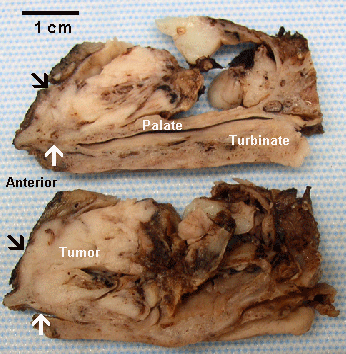
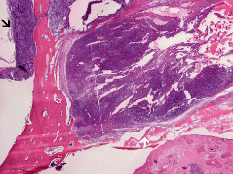
The two images on the left are sagittal sections of the decalcified maxillectomy specimen. The white arrow points to the location where the hard palate is invaded. The black and white arrows locate the areas with bone invasion and corresponding locations are shown in the histologic image on the right.