Calretinin
CK 5/6
Inhibin
| A 30 year-old Woman with
Bilateral Tubal Ligation. October, 2007, Case 710-1. Home Page |
Cheng Z. Liu, M.D., Ph.D., Kar-Ming Fung, M.D., Ph.D. Last update: December 1, 2007.
Department of Pathology, University of Oklahoma Health Sciences Center, Oklahoma City, Oklahoma
Clinical information: The patient was a 30 year-old woman who was admitted for bilateral tubal ligation. Her medical history and physical examination was unremarkable. The following images are taken from one of her excised portion of fallopian tube.
|
|
|
|
|
|
|
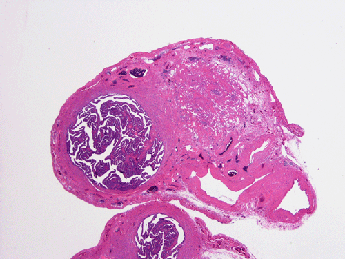
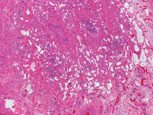
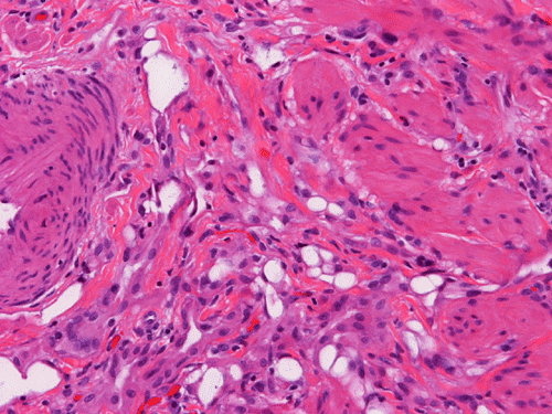
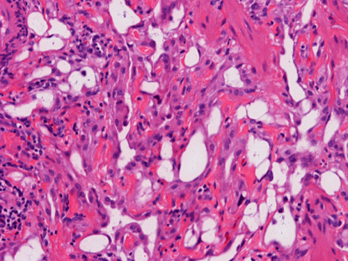
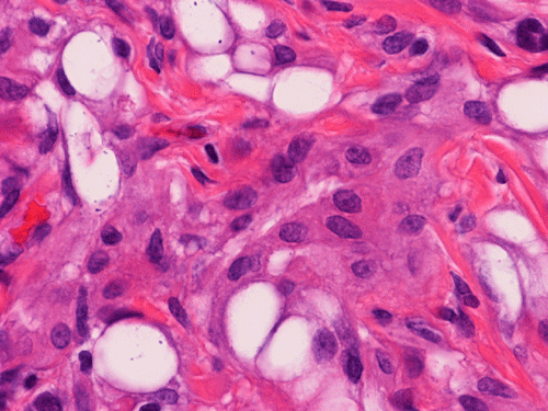
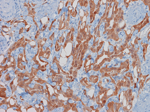
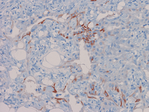
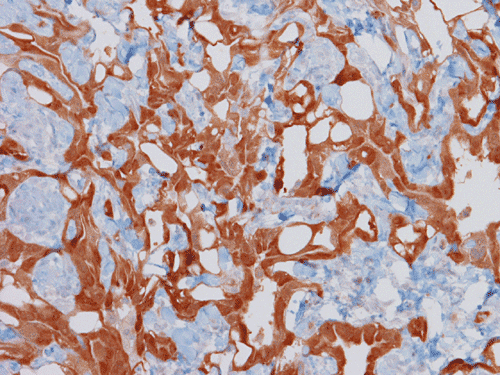
| A. | B. | C. | D. | E. | |
|
|
|
|
|||
|
F. Calretinin |
G. CK 5/6 |
H. Inhibin |
Pathology of the Case:
On scanning magnification, there is a round lesion in tissue around the fallopian tube (arrows in Panel A). The lesion does not invade into the tube nor distort or compress the tube. The cross sectional dimension is about the same diameter of the fallopian tube. On low and medium magnification, the lesion is composed of a collection of small microcysts intermingled with the smooth muscle bundles of the wall of the fallopian tube (Panel B and C). On higher magnification, the cyst or gland like spaces are lined by epithelioid to spindle cells with bland nuclei and an abundant amount of amphophilic cytoplasm (Panel D and E). The spindle cells are positive for calretinin, cytokeratin 5/6, and inhibin (Panel F, G, and H).
| DIAGNOSIS: Adenomatoid tumor of fallopian tube. |
Discussion:
General Information:
Adenomatoid tumor is a benign tumor of mesothelial origin and it can be seen in multiple organs that have mesothelial lining. They are usually seen in the genital organs including the fallopian tube, uterus, testes, and ovary. Some unusual cases have been described in the adrenal glands and small intestine 1, 2, 3. Adenomatoid tumor is the most common benign tumor of the fallopian tube. Most of these cases are incidental findings during autopsy or removal of the fallopian tubes for other purposes in middle aged or elderly woman. Most of the adenomatoid tumors are small, in the range of 1-2 cm, and mostly asymptomatic. Larger tumors may cause symptoms.
Pathology:
Macroscopically, ademonatoid tumors are well-circumscribed nodules with a gray-white cut surface. With the mesothelial origin, it would not be surprised that they are typically found in a subserosal location.
Histologically, the salient feature is small, microcystic or gland-like cystic spaces lined by flattened cells. Some of the tumor cells may arrange in cords and tubules. Hyaluronic acid rich material that can be easily demonstrated by Alcian blue stain are present in the small glands and cysts. The tumor cells are medium in size and contain moderate to abundant amphophilic to eosinophilic cytoplasm. Prominent cytoplasmic vacuoles that suggest signet ring cells may be present. This is an important feature to know in order not to confuse these tumors with primary and metastatic carcinomas particularly when the specimen is obtained from the ovary. Although these tumor appears well-circumscribed on gross examination, these tumor appears infiltrative on microscopically and should not be confused with adenocarcinoma. The key to avoid this pitfall is that adenomatoid tumor have bland nuclear feature and no mitotic figures. Infaction can occur in adenomatoid tumor.
Immunohistochemically, adenomatoid tumor is positive of cytokeratin. This may confuses their distinction from adenocarcinoma further. Because of their mesothelial orign, they are positive for calretinin, cytokeratin 5/6, and WT-1. These tumors are negative or at most weakly positive for carcinomembryonic antigen (CEA), Ber-EP4, and B72.3. As demonstrated in this case, they can be positive for inhibin. These featues help to differentiate adenomatoid tumor from adenocarcinoma.
Reference: