Elastic
Elastic
Trichrome
Trichrome
SMA
SMA
Trichrome
| A
41 year-old Man with Headache, Altered Mental Status, and a Large
Intraventricular Hemorrhage. February, 2008, Case 802-1. Home Page |
Song Lu, M.D, Ph.D.1, Kar-Ming Fung, M.D., Ph.D. 2 , Jiang Qian , M.D., Ph.D. 1 Last update: March 31, 2008.
1Department of Pathology, Albany Medical College, Albany, NY and 2Department of Pathology, University of Oklahoma Health Sciences Center, Oklahoma City, Oklahoma
Clinical information:
The patient was a 41 year-old Hispanic man. One of his friends found him lying in a puddle of blood in a day in May. According to the other information, he had a history of seizure and had been complaining of headache. He started vomiting blood and became unresponsive. He was admitted to the hospital by the EMS.
He lived in a shelter and there was no knowledge of any past medical history, medication, or other details regarding the patient at the time of admission. On physical examination upon admission to the emeregency room, there was dried blood in his nose and his mouth. He was slightly hypertensive and tachycardic. His lung was clear on auscultation and his chest movement was symmetrical. A nasogastric tube was placed and recovered about 200 ml of coffee-ground substance. A CT scan of his head demonstrated a large intraventricular hemorrhage that involved all of the ventricles. The clinical impression was a possible rupture of a distal pericallosal aneurysm. He was sedated and treated for hypertension and possible aspiration pneumonia. Bilateral ventriculostomies were placed in the emergency room.
The patient developed low grade fever in the next several days and his temperature started to spike to around 103 °F about 5 days after admission. Laboratory examination at that time showed elevated WBC, with 64% neutrophils, anemic with Hb 8.4. His CSF cultures were negative for bacteria but his endotrachial aspirate was tested positive for Hafnia alvei (Gram-negative rods). He continued to have tachycardia, tachypnea, hyperthermia, and could not be weaned off from the ventilator. Limited family history was subsequently established, which was significant for intracranial bleedings in several other family members. His condition continued to deteriorate and he developed metabolic acidosis, hypernatremia, hyperkalemia, elevated ALT and AST, markedly elevated creatine kinase, decreased GFR, acute renal failure, and DIC-like coagulation profile on June 2. Both systolic blood pressure and heart rate dropped to the 60s. Despite all possible measures taken, there was no significant improvement on the patient’s condition. On the 19th hospital day, the patient was made DNR and he expired later that day. An autopsy limited to the brain was performed.
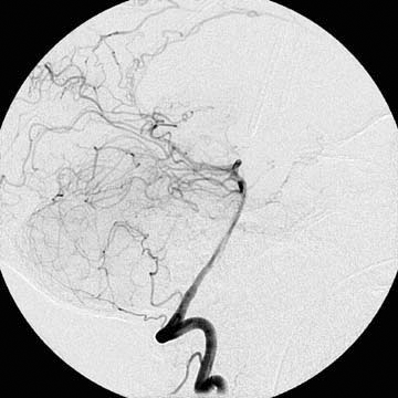
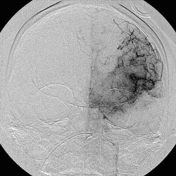
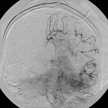
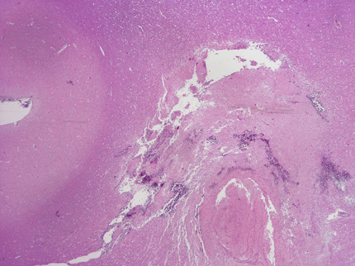
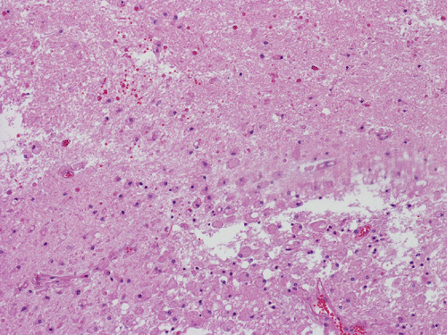
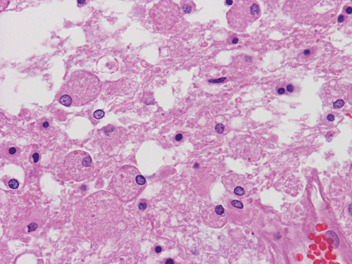
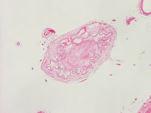
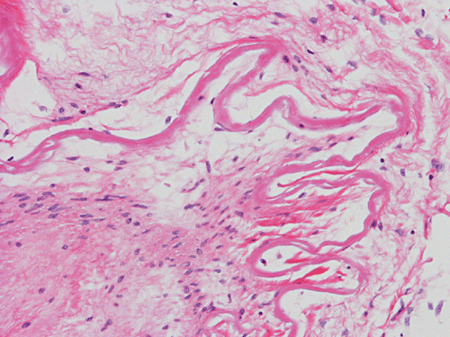
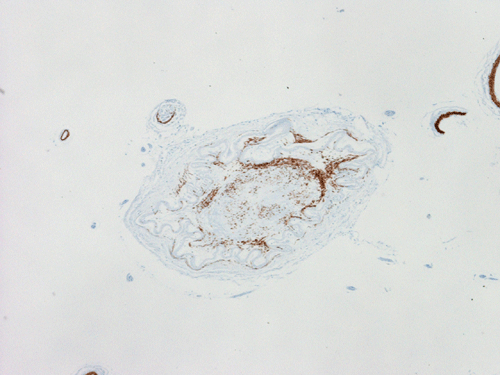
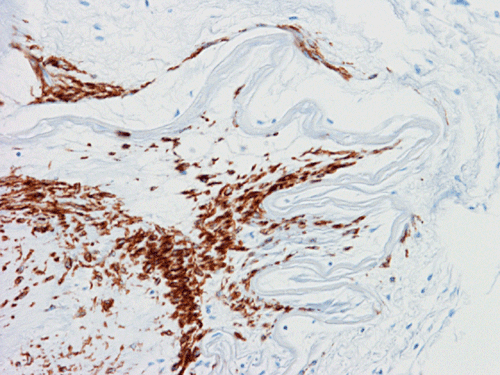
A diagnostic angiography was done during his hospitalization and yielded the following images.The followings are representative images from autopsy. Panel D to E are taken from the softened area of the right frontal lobe. Panel G and H are taken from the blood vessels of the circle of Willis. SMA stands for immunohistochemistry for smooth muscle actin.
|
|
|
|
|
|
|
| A | B | C | D | E | F |
|
|
|
|
|
|
|
| G | H |
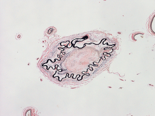
I. Elastic |
J. Elastic |
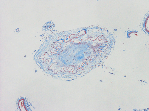
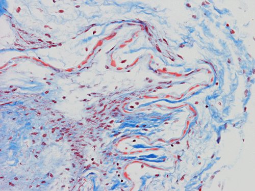
K. Trichrome |
L. Trichrome |
|
|
|
|
|
||
|
M. SMA |
N. SMA |
Scanne slide |
Scanned slide Trichrome |
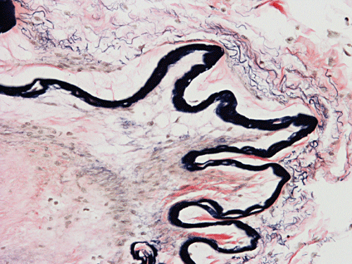
Pathology of the case: The abnormal circulation and hazy, or "puff of smoke" like abnormal vasculature is best demonstrated with the angiogram (Panel A, B, and C). On gross examination, the lateral ventricles are filled and dilated with blood. There are some softening of brain tissue around the hemorrhage and in the frontal area (Panel D, E, and F). Vessels at circle of Willis appears whitish but not dilated. On cross section, the lumen is poorly defined or collapsed. On histological sections stained with hematoxylin and eosin stain, the vessel has a wavy internal elastic lamina. The intima has thickened to the point that the lumen is almost complete occluded. There is no signifcant inflammatory cell or histiocytic infiltration (Panel G and H). On elastic stain (Panel I and J), the internal elastic lamina is intact and without duplication or splitting. Substantial deposition of collagenous fibrous tissue is demonstrated by Masson's Trichrome (Panel K and L). Immunohistochemistry for smooth muscle actin (SMA) demonstrated a large amount of smooth muscle in the thickened intima (Panel M and N).
| DIAGNOSIS: Moyamoya disease associated with massive intraventricular hemorrhage. |
Discussion:
General Information
In Japanese, the term moyamoya means a puff of smoke drifting in air . Moyamoya disease or moyamoya syndrome refer to an angiographical picture of hazy (or a puff of smoke) like appearance of collateral circulations that are formed due to vascular occlusion involving the circle of Willis. The term moyamoya syndrome covers both primary moyamoya disease and cases of angiographic moyamoya appearance associated with a variety of different diseases. Cases that do not fulfill the full criteria of moyamoya diseases, such as the unilateral cases are termed probable moyamoya disease. Moyamoya disease was first described in Japanese but it is nows known that neither moyomoya disease nor moyamoya syndrome are limited to Japanese or Asian. Even in Japan, however, it is not a common disease and has an incidence of 0.35 per 100,000 population 1. There is an overall slight female preponderance. Suzuki and Kodama classified the changes 2, 3 by conventional angiographic method. A recent studies using magnetic resonance angiography and a 0-10 scoring system generated similar results as compared to the conventional system proposed by Suzuki and MRA has the advantage of being a non-invasive procedure 4.
The etiology of moyamoya disease and moyamoya syndrome is not clear. However, they share the common mechanism of occlusion of the artery in circle of Willis. The occlusion of the internal carotid artery is a slow and progressive process that lead to the formation of numerous dilated, thin-walled collateral arteries, the foundation of the characteristic hazy, smoke like angiogram. In children, the clinical manifestations are typically due to cerebral ischemia and the most common manifestation is alternating hemiparesis. In adult, hemorrhage from the thin-walled blood vessel as illustrated in this case is the most common feature.
The etiology in primary moyamoya disease is uncertain. Most of these cases are seen in children or adolescence and are not associated in known conditions. Moyamoya diseases associated with a variety of systemic diseases including Graves disease 5, Down syndrome 6, beta-thalassemia 7,sickle cell disease 8, AIDS 9, and others have been described. Familial cases have also been described. Although the clinical manifestations of familial cases appear to be similar to sporadic cases 10, these cases may carry specific genetic alterations 11, 12. A candidate gene has been identified on chromosome 3p24.2-p26 12. Moyamoya syndrome has also bee associated with a consortium of inherited conditions including neurofibromatosis-1, tuberous sclerosis, Marfan's syndrome, Alpert's syndrome and others.
Pathology:
The salient features are bilateral stenosis or occlusion of the internal carotid arteries. The stenoic or occluded portion may appear stiff, fibrotic, or whitish due to the fibrotic thickening. The stenosis or occlusion usually spans from the distal portions of the internal carotid artery towards the anterior and middle cerebral arteries. These occlusions or stenoses lead to the formation of numerous collateral arteries which would form an irregular vascular network on the pial surface. These vessels would also penetrate the brain parenchyma. These vessels are typically dilated and thin-walled and they branch from the posterior portion of the circle of Willis.
Histologically, there is dramatic thickening of the intima with preservation of the internal elastic lamina. Atherosclerotic changes are not typically present. The internal elastic lamina becomes extremely wavy and may appear double-barreled (duplicated) or triplicated appearance. Inflammatory cells are not usually present. Thrombosis or fibrotic occlusion of the lumen can also occur. In an earlier study 12, vessels showing rupture ranged from 50 to 530 microns in diameter; they were dilated, some had fibrin deposits in the wall, fragmented elastic laminae and attenuated media. Non-ruptured perforating arteries (diameter 200 to 550 microns) revealed microaneurysm formation, focal fibrin deposits and marked attenuation of the wall thickness with diminution of the elastic lamina. In another studies, smooth muscle cells, macrophages and T-cells have been demonstrated in the thickened intima 14. In a single case study, immunohistochemistry has not demonstrated vascular endothelial growth factor (VEGF) or VEGF receptor in the collateral vessels 15.
Reference:
Kuriyama S, Kusaka Y, Fujimura M, Wakai K, Tamakoshi A, Hashimoto S, Tsuji I, Inaba Y, Yoshimoto T. Prevalence and clinicoepidemiological features of moyamoya disease in Japan: findings from a nationwide epidemiological survey. Stroke. 2008 39:42-7.
Suzuki J, Takaku A. Cerebrovascular "moyamoya" disease. Disease showing abnormal net-like vessels in base of brain. Arch Neurol. 1969 20:288-99.
Suzuki J, Kodama N. Moyamoya disease--a review. Stroke. 1983 14:104-9.
Houkin K, Nakayama N, Kuroda S, Nonaka T, Shonai T, Yoshimoto T. Novel magnetic resonance angiography stage grading for moyamoya disease. Cerebrovasc Dis. 2005 20:347-54.
Hsu SW, Chaloupka JC, Fattal D. Rapidly progressive fatal bihemispheric infarction secondary to Moyamoya syndrome in association with Graves thyrotoxicosis. AJNR Am J Neuroradiol. 2006 27:643-7.
Bhalala US, Parekh PR. Moyamoya syndrome in a child with Down syndrome. Indian J Pediatr. 2005 72:635-7.
Marden FA, Putman CM, Grant JM, Greenberg J. Moyamoya disease associated with hemoglobin Fairfax and beta-thalassemia. Pediatr Neurol. 2008 38:130-2.
Dobson SR, Holden KR, Nietert PJ, Cure JK, Laver JH, Disco D, Abboud MR. Moyamoya syndrome in childhood sickle cell disease: a predictive factor for recurrent cerebrovascular events. Blood. 2002 99:3144-50.
Sharfstein SR, Ahmed S, Islam MQ, Najjar MI, Ratushny V. Case of moyamoya disease in a patient with advanced acquired immunodeficiency syndrome. J Stroke Cerebrovasc Dis. 2007 16:268-72.
Seol HJ, Wang KC, Kim SK, Hwang YS, Kim KJ, Cho BK. Familial occurrence of moyamoya disease: a clinical study. Childs Nerv Syst. 2006 22:1143-8.
Ikeda H, Yoshimoto T. Specific genetic characteristics in patients with familial moyamoya disease. J Stroke Cerebrovasc Dis. 2005 14:244-50.
Ikeda H, Sasaki T, Yoshimoto T, Fukui M, Arinami T. Mapping of a familial moyamoya disease gene to chromosome 3p24.2-p26. Am J Hum Genet. 1999 64:533-7.
Yamashita M, Oka K, Tanaka K. Histopathology of the brain vascular network in moyamoya disease. Stroke. 1983 14:50-8.
Masuda J, Ogata J, Yutani C. Smooth muscle cell proliferation and localization of macrophages and T cells in the occlusive intracranial major arteries in moyamoya disease. Stroke. 1993 24:1960-7.
Takekawa Y, Umezawa T, Ueno Y, Sawada T, Kobayashi M. Pathological and immunohistochemical findings of an autopsy case of adult moyamoya disease. Neuropathology. 2004 24:236-42.
Cases of the Month Evaluation Coordinator: KarMing-Fung@ouhsc.edu
Copyrights reserved.