| A 59 year-old Woman with a
Prolonged History of Diarrhea for a Few Months. April, 2008, Case 804-1. Home Page |
Jinous Saremian, M.D. 1, Matthew M. Yeh, M.D., Ph.D.2 Last update: June 1, 2008.
1 Department of Pathology, University of Oklahoma Health Science Center, Oklahoma City, Oklahoma and 2 Department of Pathology, University of Washington School of Medicine, Seattle, WA.
Clinical information: The patient was a 59 year-old woman with watery diarrhea for 4 months. The mucosa is unremarkable on endoscopic examination. There was no ulcer or friable area. A biopsy was obtained from the colon and yielded the following images.
|
|
|
|
|
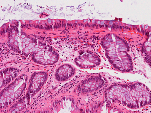
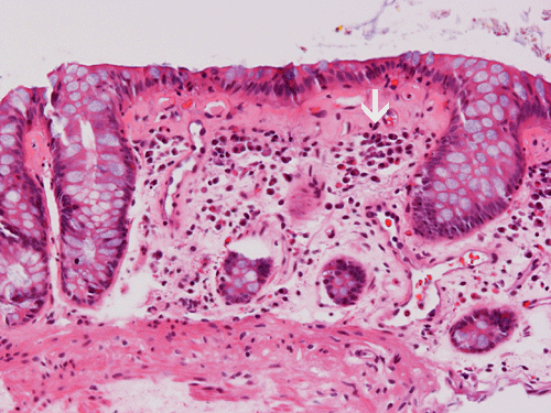
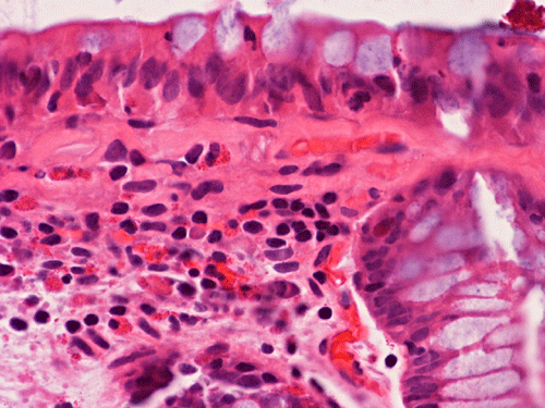
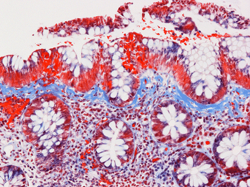
| A. | B. | C. | D. |
Pathology of the Case:
The salient feature of the biopsy specimen is a thickened band of eosinophilic, collagen-like material under the epithelium associated with cell infiltration composed predominantly of lymphocytes and plasma cell (Panel A). The inflammatory cell infiltration is not particularly intense but has a "top heavy" type of distribution that accentuates in the upper portion of the lamina propria (arrow in Panel B). No polymorphonuclear leukocytic infiltration to indicate active inflammation is noted (Panel C). On Masson's trichrome stain, the thickend subepithelial layer appears positive for collagen, confirming significantly thickened subepithelial collagen table (Panel D).
| DIAGNOSIS: Collagenous colitis. |
Discussion:
General information
Microscopic colitis is a disease of chronic watery diarrhea with microscopic changes in the colonic mucosa yet without significant abnormalities on colonoscopy. Collageneous colitis (CC) is a type of microscopic colitis and is histologically characterized by a thickened subepithelial collagen layer, “top heavy” prominent chronic inflammation in the lamina propria, and increased intraepithelial lymphocytes in the colonic mucosa with the clinical manifestations of chronic, non-bloody watery diarrhea, macroscopically normal-appearing colonic mucosa without radiological abnormalities 1. It is far more common in women, with a female to male ratio of 6-8:1 and a median age of diagnosis at 59 years. Approximately 40% of patients have an associated disease and the most common of these include rheumatoid arthritis, thyroid disorders, celiac disease, and diabetes mellitus. Certain drugs including non-steroidal anti-inflammatory drugs (NSAIDS) and autoimmune diseases have been suggested to be associated with CC 2.
As microscopic colitis has normal appearing mucosa on colonoscopy, a single biopsy from only one site may not be able to show the pathologic changes. It is thus important to take multiple biopsies at different location of the colon in order to establish a diagnosis of CC. The use of inflammatory markers in stools has been suggested as a non-invasive tool in the diagnosis of CC 3. Although CC is typically seen in adult, rare cases have been reported in the pediatric age group 4.
Pathology:
In normal colon, a delicate subepithelial collagen table less than 3µm in thickness is visible just beneath the luminal epithelium. In collagenous colitis, there is abnormal deposition of collagen immediately beneath the basement membrane. This collagen deposition forms a subepithelial band in the superficial lamina propria, which is readily recognizable as a layer of eosinophilic, irregular deposition by hemtoxylin and eosin stain. These abnormal depositions stain positive for collagen on Masson’s trichrome stain. In addition, the thickened subepithelial collagen table incorporates capillaries and inflammatory cells.
By immunohistochemical staining, this thickened band contains collagens type I, III, and VI as well as tenascin. Immunohistochemistry for basement membrane components, such as type IV collagen and laminin, do not highlight this layer, further confirming that this deposition is collagen rather than basement membrane in nature. On electron microscopy, this layer of collageneous deposition is separated from and beneath the basement membrane. In a same patient, the thickness of this collagenous band can vary throughout the colon and the transverse colon generally has the thickest deposition. The rectum can lack a thick subepithelial collagen band in up to 33% of cases, and in a small percentage of patients, the rectum can be histologically normal with no increase in inflammation. Consequently, multiple biopsy specimens should be taken in areas proximal to the rectosigmoid colon if the clinical suspicion of collagenous colitis is strong 1.
The lamina propria is typically expanded by a mixture of inflammatory cells, including plasma cells, lymphocytes, eosinophils and mast cells. These cells tend to have a “top heavy” pattern and more inflammatory cells are found immediately under the collagenous band than in deep portion of the lamina propria. The other distinctive component of collagenous colitis is increased intraepithelial lymphocytes. The intraepithelial lymphocytes of collagenous colitis are predominantly CD8 cells which express the alpha- beta form of T cell receptor. Surface epithelial damage (flattening, detachment) may also be present 1 but these are non-specific features as they are also present in other form of colitis.
Pathogenesis:
The cause of collagenous colitis is largely unknown. Hypotheses for the etiology of collagenous colitis include immune dysregulation, abnormalities in pericryptal fibroblasts, intraluminal bacterial agents or toxins, and drug-induced damage. A plethora of inflammatory mediators have been found to be elevated in collagenous colitis with the most recently investigated including nitric oxide, and vascular endothelial growth factor 1. The mechanisms of diarrhea are variable between patients. Fasting improves, but would not totally abate the diarrhea in most patients, suggesting both an osmotic and a secretory component to the diarrhea.
Differential diagnosis:
Lymphocytic Colitis: There are many overlapping features between lymphocytic colitis and CC. They share similar clinical manifestations with negative endoscopic findings. The histologic similarities include increased intraepithelial lymphocytes, surface epithelial damage, and increased chronic inflammatory cells within the lamina propria. In contrast to collagenous colitis, there is no increased thickness of the subepthelial collagen band in lymphocytic colitis..
Brainerd Diarrhea: The term Brainerd Diarrhea has been applied to outbreaks of diarrhea of unknown etiology that is characterized by acute onset and prolonged duration. Unlike typical infectious diarrhea, chronic watery diarrhea develops with symptoms lasting longer than 6 months and often for years. Colonic biopsy specimens reveal surface epithelial lymphocytosis without distortion of the mucosal architecture, surface degenerative changes, or a thickened subepithelial collagen plate.
References: