Von Kossa
Von Kossa
| A 52 year-old Woman with
Diabetes, Renal Failure, and Abdominal Pain. September, 2008, Case 809-2. Home Page |
Colin C. Pritchard, M.D., Ph.D., Matthew M. Yeh, M.D. Ph.D.
Department of Pathology, University of Washington School of Medicine, Seattle, WA. Last update: August 27, 2008.
Clinical information: The patient was a 52-year old woman who presented with a chief complaint of abdominal pain of two weeks duration. She had a history of type II diabetes mellitus resulting with end-stage renal disease which required ongoing dialysis. Laboratory studies revealed a serum calcium level of 11.2 mg/dL (reference range 8.9 – 10.2 mg/dL) and a phosphate level of 5.5 mg/dL (reference range for adult females 2.5 – 4.5 mg/dL). Upper gastrointestinal endoscopy showed a granular, nodular mucosa in the gastric body and fundus with a patchy distribution.
|
|
|
|
|
|
|
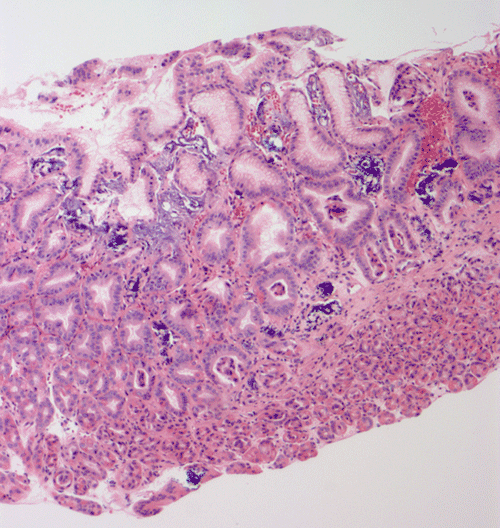
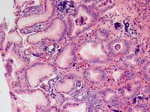
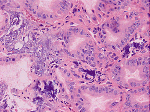
| A. | B. | C. |
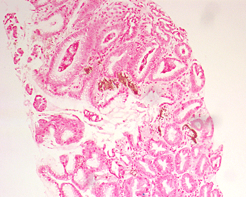
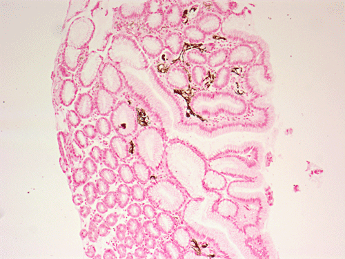
D. Von Kossa |
E. Von Kossa |
Pathology of the case: At low power magnification, the biopsy appears to be a portion of gastric fundic-type mucosa (Panel A). Prominent scattered irregular, amorphous basophilic substances are present in small deposits in the superficial lamina propria, abutting the gastric epithelium (Panel A, B, and C). The mucosa has a reactive appearance, with enlarged nuclei, prominent nucleoli and regenerative appearing glands (best seen in Panel C). An iron stain is negative on these deposits arguing against that these are iron containing substances, hemosiderin or other hemoglobin breakdown products (Panel D). These irregular, basophilic, amorphous material is positive by von Kossa stain (Panels E), consistent with calcium depositions. There is no evidence of intestinal metaplasia, dysplasia, or neoplasia. No significant foveolar hyperplasia or inflammation is present.
| DIAGNOSIS: Gastric mucosal calcinosis. |
Clinical follow up on this case: Serum calcium and phosphate levels remained persistently elevated and the patient developed systemic medial calcifications of arteries (calciphylaxis) with associated wounds on the upper extremities and abdomen. Parathyroidectomy was performed 5 months after the current biopsy for secondary hyperparathyroidism. This helped normalize calcium and phosphate, but the patient unfortunately died one year later from complications of long-standing coronary artery disease, possibly compounded by calciphylaxis. An autopsy was not performed.
Discussion:
General Information
Gastric mucosal calcinosis is a rare finding in mucosal biopsies and is most frequently associated with conditions which cause hypercalcemia and hyperphophatemia 1. Although gastric mucosal calcinosis is often an incidental biopsy finding, it is important to report as a potential heralding sign of systemic calcinosis, which can be fatal if critical organs such as the heart are involved 2. Patients with end-stage kidney disease or solid organ transplant are particularly prone to gastric mucosal calcinosis 3, 4, 5. There is also limited evidence that medications such as aluminum-containing antacids and sucralfate are associated with gastric mucosal calcinosis 4, 6. The alkaline environment in the superficial gastric lamina propria presumably serves a preferential site for calcium deposition in systemic conditions which alter the serum biochemical milieu.
Incidence
The incidence of gastric mucosal calcinosis in the general population is unknown. In small cohorts of patients with chronic renal disease the incidence has been reported between 13-60%, with the highest incidence in patients with chronic uremia who were on dialysis 2, 6, 7. In one study of solid organ transplant patients, 18/55 (33%) had gastric mucosal calcinosis compared with 3/59 (5%) of a control group with gastric ulcers 4. Gorospe et. al. reported the incidence of gastric mucosal calcinosis as less than 0.1% of routine gastric biopsies collected in their sizable upper GI pathology service 1.
Classification, etiology, and pathogenesis
Gastric mucosal calcinosis has been classified as either metastatic, dystrophic, or idiopathic 1, 3. In metastatic calcification, the most common cause of calcinosis is calcium salts deposit in normal gastric tissue as a result of altered serum electrolytes: classically hyperphosphatemia and hypercalcemia. Diseases associated with metastatic calcification include end stage kidney disease 2, 8, 9, primary hyperparathyroidism 7, 10, multiple myeloma 11, 12, metastatic malignancy, hypervitaminosis D 7, 13, and the tumor lysis syndrome 14. Serum elevations of calcium and phosphate result in increased formation of insoluble calcium phosphate salts 9. The solubility of calcium phosphate is inversely related to pH, resulting in deposition of calcium precipitates in the relative alkaline conditions of the superficial gastric lamina propria, as well as in the kidneys, lungs, and left cardiac chamber 1, 7.
In dystrophic calcification, calcium salts deposit as a result of tissue damage, typically from inflammation, fibrosis, or trauma. Dystrophic calcifications may be seen in association with gastric ulcers, or malignancy in the setting of a normal serum calcium and phosphate.
Clinical Presentation
Gastric mucosal calcinosis is not typically associated with symptoms, although there are rare case reports of dyspepsia and epigastric pain that could not be explained by other causes 14, 15. Thus, gastric calcinosis will most commonly be an incidental finding on biopsy. As mentioned above, it is important for the pathologist to report the finding in biopsies because of the association with metastatic calcification in critical organs.
Diagnosis: Endoscopic and histopathologic features
Endocopic findings are typically 1 -5 mm flat white plaques or nodules seen diffusely throughout the stomach 16. Plaques may become confluent in areas, mimicking the appearance of a gastric ulcer 14. Microscopically, amorphous basophilic foreign material is seen in the superficial lamina propria just underlying the epithelium in any part of the stomach 1, 2, 4, 5, 15. Deposits may also be seen deeper in the lamina propria or associated with the muscularis mucosa. In more severe cases, calcium deposits may be associated with submucosal vessels causing luminal stenosis 2. Linear deposition of calcium salts can be seen in association with the basement membrane. The deposits do not show birefringence to polarized light 6, 15. There may be histiocytes lining calcium deposits or without significant inflammation. Greenson et. al. 4 described a subset of cases in which there were deposits of amorphous refractile material which was only sparsely calcified at the periphery.
While there is often no associated pathology with gastric mucosal calcinosis, there are reports of associated reactive gastropathy 4, chronic active gastritis 6, foveolar hyperplasia 4, 6, atrophic gastritis 16, ulceration 4, and mucosal edema 4. It is unclear how significant the association with gastric mucosal calcinosis is with any of these conditions. There is no evidence that Helicobactor pylori infection is associated with gastric mucosal calcinosis 1.
References:
Gorospe M, Fadare O. Gastric mucosal calcinosis: clinicopathologic considerations. Adv Anat Pathol 2007, 14:224-228.
Kuzela DC, Huffer WE, Conger JD, Winter SD, Hammond WS. Soft tissue calcification in chronic dialysis patients. Am J Pathol 1977, 86:403-424.
Castaigne C, Martin P, Blocklet D. Lung, gastric, and soft tissue uptake of Tc-99m MDP and Ga-67 citrate associated with hypercalcemia. Clin Nucl Med 2003, 28:467-471.
Greenson JK, Trinidad SB, Pfeil SA, Brainard JA, McBride PT, Colijn HO, Tesi RJ, Lucas JG. Gastric mucosal calcinosis. Calcified aluminum phosphate deposits secondary to aluminum-containing antacids or sucralfate therapy in organ transplant patients. Am J Surg Pathol 1993, 17:45-50.
Munoz SJ, Nagelberg SB, Green PJ, Angstadt JD, Yang SL, Jarrell BE, Maddrey WC. Ectopic soft tissue calcium deposition following liver transplantation. Hepatology 1988, 8:476-483.
Stroehlein KB, Stroehlein JR, Kahan BD, Gruber SA. Gastric mucosal calcinosis in renal transplant patients. Transplant Proc 1999, 31:2124-2126.
Mulligan R. Metastatic Calcification. Archives Pathology 1911, 43:177-230
Milliner DS, Zinsmeister AR, Lieberman E, Landing B. Soft tissue calcification in pediatric patients with end-stage renal disease. Kidney Int 1990, 38:931-936.
Parfitt AM. Soft-tissue calcification in uremia, Arch Intern Med 1969, 124:544-556.
Hwang GJ, Lee JD, Park CY, Lim SK. Reversible extraskeletal uptake of bone scanning in primary hyperparathyroidism. J Nucl Med 1996, 37:469-471.
Esser JP, Oei HY, Kwekkeboom DJ, Krenning EP. Diffuse lung and stomach uptake of Tc-99m oxidronate (HDP). Clin Nucl Med 2003, 28:845-846.
Reitz MD, Vasinrapee P, Mishkin FS. Myocardial, pulmonary, and gastric uptake of technetium-99m MDP in a patient with multiple myeloma and hypercalcemia. Clin Nucl Med 1986, 11:730.
Corstens F, Kerremans A, Claessens R. Resolution of massive technetium-99m methylene diphosphonate uptake in the stomach in vitamin D intoxication. J Nucl Med 1986, 27:219-222.
Avci Z, Alioglu B, Canan O, Ozcay F, Celasun B, Sarialioglu F, Ozbek N. Calcification of the gastric mucosa associated with tumor lysis syndrome in a child with non-Hodgkin lymphoma. J Pediatr Hematol Oncol 2006, 28:307-310.
Saab S, Venkataramani A, Behling CA, Savides TJ. Gastric mucosal calcinosis in a patient with dyspepsia. J Clin Gastroenterol 1996, 22:156-164.
Ou Tim L, Hurwitz S, Tuch P. The endoscopic diagnosis of gastric calcification. J Clin Gastroenterol 1982, 4:213-215.