Squash
Squash
Frozen
Frozen
Frozen
| A 65 year-old Woman with an
Intraventricular Mass. January, 2005, Case 501-2. Home Page |
Brandon Guthery, B.S. (MS-IV)1, Kalliopi Petropoulou, M.D.2, Kar-Ming Fung, M.D., Ph.D.3 Last update: February 12, 2005.
1 Fourth year medical student, Class of 2005, College of Medicine, University of Oklahoma, 2 Department of Radiology and 3 Department of Pathology, University of Oklahoma Health Sciences Center, Oklahoma City, Oklahoma
Clinical information: The patient was a 65 year-old woman who sustained a minor automobile accident. An MR imaging scan was performed and an unexpected lesion was demonstrated. The lesion was removed. The followings are the selected MR images and the representative histologic photos.
|
|
|
|
|
|
|
| A. | B. | C. |
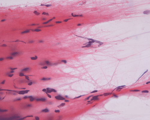
D. Squash |
E. Squash |
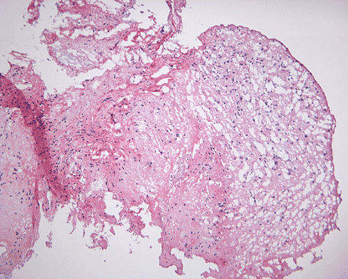
F. Frozen |
|
|
|
|
|
|
|
|
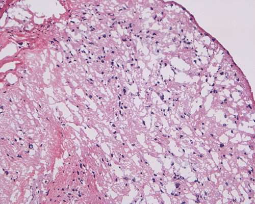
G. Frozen |
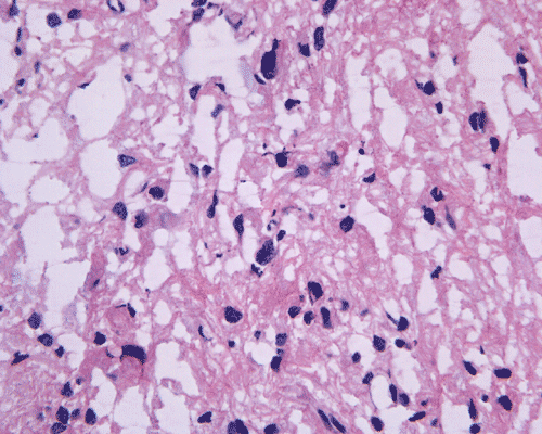
H. Frozen |
I. | J. | K. | L. |
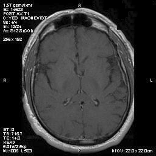
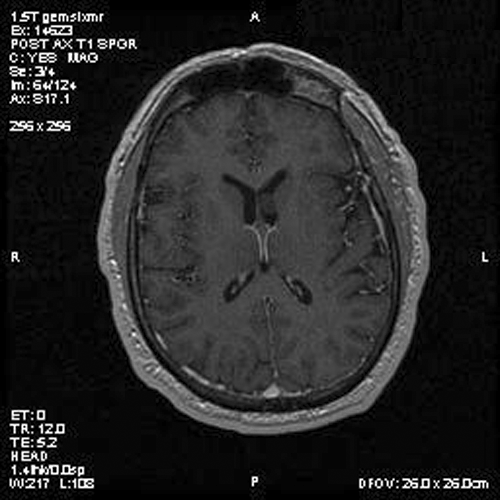
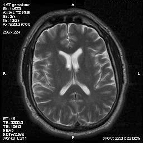
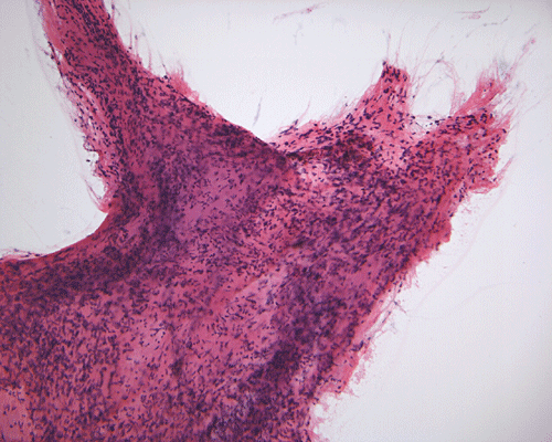
Panel A is T1-weighted post contrast MR image, Panel B is a SPGR T1-weighted post contrast MR image and Panel C is FSE T2-weighted image. Panel D and E are taken from cytologic preparation from intraoperative consultation. Panel F to H are taken from frozen sections. Panel I to L are taken from paraffin section.
Image of the case: In the left frontal horn there is a well demarcated, subependymal mass measuring approximately 1.1 x 0.5 cm which is relatively hyperintense to white matter on T1-weighted image (Panel A and B) as well as T2-weighted image (Panel C) sequences. It does not enhance on the post contrast images. There is no obstruction of the foramen of Monro.
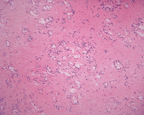
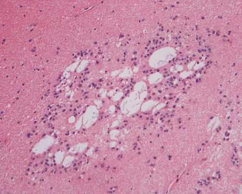
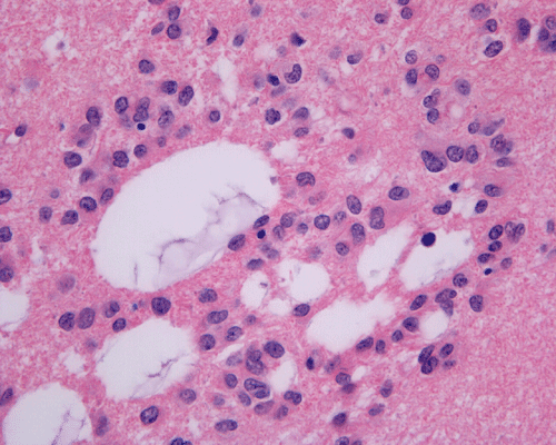
Pathology of the case: A small specimen was obtained by endoscopic biopsy. The specimen cannot be smear out after being squashed and remain as several, large, stellate-shaped cohesive clumps (Panel D). The edges of these clumps are relatively thin and allows better observations. The nuclei do not appear to be pleomorphic. Many elongated cytoplasmic processes can be seen (Panel E) and are suggest a glial nature of these cells. The lesion gives a spongy appearance on low-magnification (Panel F). On medium-magnification, there are some clustering of nuclei (Panel G). On high-magnification, the nuclei appear hyperchromatic and pointy but no substantial pleomorphism, prominent nucleoli, or mitotic figure. Some bluish, mucoid material is identified in some microcysts (Ú in Panel H).
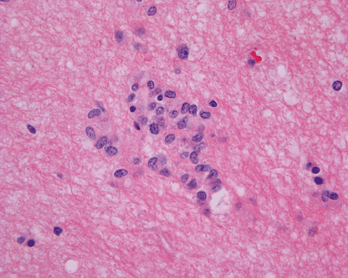
There is no endothelial proliferation or necrosis. An intraoperative diagnosis of glial neoplasm was made. The lesion was entirely resected. On paraffin section, the lesion has a hypocellular background decorated by many small microcysts, often in clusters, that contain mucoid material and small cluster of nuclei that resemble bundles of flowers (Panel I and J). The nuclei are bland. There was no mitosis, endothelial proliferation or necrosis (Panel K and L).
| DIAGNOSIS: Subependymoma, WHO grade I/IV. |
Discussion: General Information Pathology Prognosis
General Information
Subependymomas are composed of glial tumor cell clusters embedded in an abundant fibrillary matrix with frequent microcystic change. It is a histologic grade I tumor in the WHO classification 1. It was first described by Scheinker in 1945 2. Astrocytic features at the histologic level in these tumors have led to other names such as: ‘subependymal astrocytoma’ and ‘subependymal glomerate astrocytoma’ that do not really reflect the true biologic phenotype of these tumors.
The most common site for these tumors is the fourth ventricle followed by the lateral ventricles. The third ventricle and septum pellucidum are involved less commonly. The spinal cord can be involved as well, and these lesions can present as cervical and cervico-thoracic intramedullary or extramedullary mass lesions 3.
Subependymomas occur in all age groups and both sexes, with a predilection for middle-aged and elderly males. Many of them remain asymptomatic and present as incidental clinical finding as illustrated in our case or at autopsy. When they are symptomatic, the salient clinical manifestations are resulted to the hydrocephalus caused by the tumor. Subependymomas comprised 8.3% of ependymal tumors in a study of 298 cases 4. Subependymomas may cause ventricular obstruction with raised intercranial pressure. Spontaneous intratumoral hemorrhage can occur 5. Spinal tumors manifest with motor and sensory deficits depending on location of the lesion.
Imaging
Typically, subependymoma occur as a fungating mass that protrudes into the ventricle. A mild to sometime significant hydrocephalus can be caused by these tumors. On T1-weighted images, they have intensity similar to that of the surrounding white matter. T2-weighted images usually do not demonstrate significant edema around these tumors. Some of its properties also varies according to the locations. In tumors arising in the fourth ventricle, calcification and heterogeneous contrast enhancement are common; whereas these findings are rare in subependymomas arising in the lateral ventricles 6.
Grossly, subependymomas present as firm, lobularted, well-demarcated, whitish nodules. Asymptomatic cases usually do not exceed 1.0 cm in diameter; while tumors that obstruct and cause hydrocephalus usually range from 3-5 cm in diameter 7, 8.
Histologically, the tumors are composed of a uniform population of ependymal cells embedded in a densely fibrillar glial matrix; with groups and islands of cells surrounded by acellular bands of glial fibers. Mitoses are rare or absent. Like other ependymal tumors, perivascular coronary arrangements of tumor cells (pseudorosettes) can occur. These arrangements, however, is usually not conspicuous, small, and relatively uncommon. When the amount of pseudorosette is substantial, a diagnosis of ependymoma should be considered seriously. Larger tumors, particularly those arising in the lateral ventricles, often show microcyst formation. Other features include hemosiderin deposition from prior hemorrhages, sclerotic blood vessels and calcifications 9. Subependymomas have the lowest rate of cell proliferation of ependymal tumors as evidenced by Ki67 (MIB-1) labelling 9 . PET scan study in one case also reveals exceedingly low rates of glucose metabolism and kinetic constants. The hypometabolism indicates low cellular density and slow growth 10. Ultrastructurally, astrocytic or ependymal cells are seen with transitional forms between these two appearances, demonstrating that the neoplasm is of one cell type. Large cells similar to ependymoglial precursor cells suggest that subependymomas may originate from cells of the adult subependymal layer 11.
Subependymoma may contain a small amount of ependymoma component. When the amount of ependymoma component is substantial, these tumors are best termed mixed ependymoma-subependymoma. These tumors are more likely to behave like an ependymoma which is a WHO grade II tumor. The proportion of ependymoma component that is needed to make this diagnosis is not clear cut.
Subependymal tumors are slow growing tumors. Surgical resection alone often offers a cure. Post operative radiation treatment are often unnecessary. Uncommon recurrent cases have been reported and successfully managed with radiation therapy 12, 13, 14.
Reference:
Wiestler O.D., Schiffer D. Subependymoma In: World Health Organization Classification of Tumors, Pathology and Genetics of Tumours of the Nervous System. 2000, Pg 80-81
Scheinker IM. Subependymoma (a newly recognized tumor of subependymal derivation). J Neurosurg 1945 2: 232-340
Lantos PL, Et al. Tumours of the nervous system. In: Greenfield’s Neuropathology 7th ed 2:833-834
Schiffer D, Chio A, Giordana MT, Migheli A, Palma L, Pollo B, Soffietti R, Tribolo A. Histologic prognostic factors in ependymoma. Childs Nerv Syst 1991 7:177-82.
Lindboe CF, Stolt-Nielsen A, Dale LG. Hemorrhage in a highly ascularized subependymoma of the septum pellucidum: case report. Neurosurgery 1992 31:741-5.
MV Chiechi, JG Smirniotopoulos and RV Jones. Intracranial subependymomas: CT and MR imaging features in 24 cases. American Journal of Roentgenology 1995 165:1245-1250.
Scheithauer BW. Symptomatic subependymoma. Report of 21 cases with review of the literature. J Neurosurg 1978 49:689-96.
Rath T, Sundgren P, Brahma B, Lieberman A, Chandler W, Gebarski S. Massive symptomatic subependymoma of the lateral ventricles: case report and review of the literature. Neuroradiology 2005 [Epub]
Prayson RA, Suh JH. Subependymomas: clinicopathologic study of 14 tumors, including comparative MIB-1 immunohistochemical analysis with other ependymal neoplasms. Arch Pathol Lab Med 1999 123:306-9.
Mineura K, Et al. Subependymoma of the septum pellucidum: characterization by PET. J Neurooncol 1997 32:143-7.
Moss TH. Observations on the nature of subependymoma: an electron microscopic study. Neuropathol Appl Neurobiol 1984 10:63-75.
Nishio S, Morioka T, Mihara F, Fukui M. Subependymoma of the lateral ventricles. Neurosurg Rev 2000 23:98-103.
Robert D. Ecker, Bruce E. Pollock. Recurrent subependymoma treated with radiosurgery. Stereotact Funct Neurosurg 2004 82:58-60
Sarkar C, Mukhopadhyay S, Ralte AM, Sharma MC, Gupta A, Gaikwad S, Mehta VS. Intramedullary subependymoma of the spinal cord: a case report and review of literature. Clin Neurol Neurosurg 2003 106:63-8.
Cases of the Month Evaluation Coordinator: KarMing-Fung@ouhsc.edu