Case No.: K-004 Quiz
Diagnosis: Chromophobe renal cell carcinoma, eosinophilic variant
Organ: Kidney
Last Updated: 12/21/2011
|
|
|
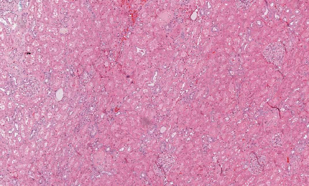
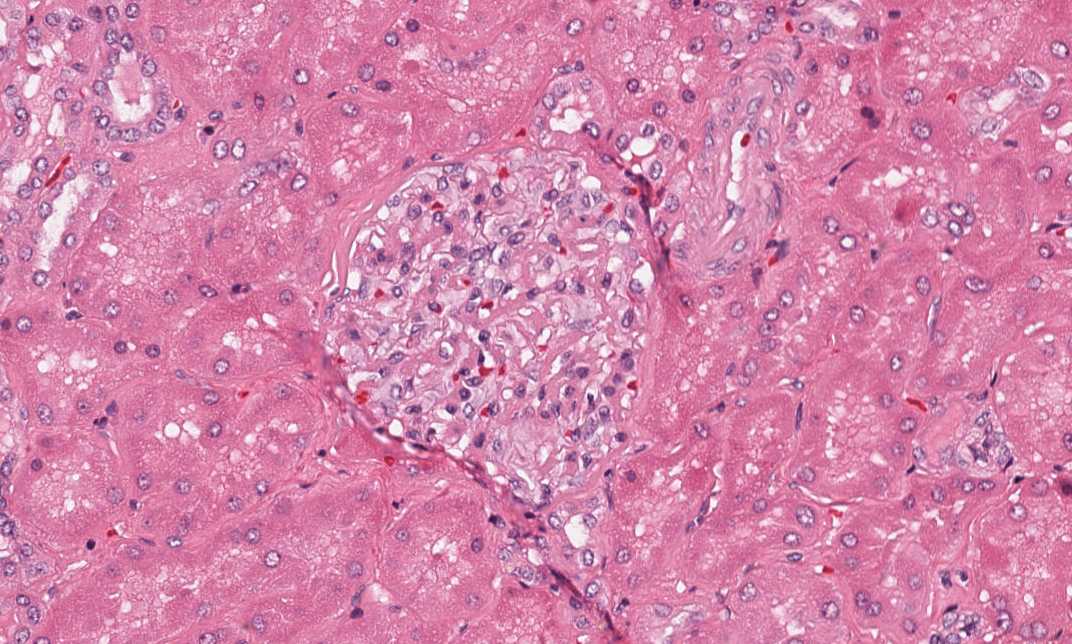
Hematoxylin & eosin |
Area 1: This image is taken from the residual renal parenchyma and many glomeruli are present. |
|
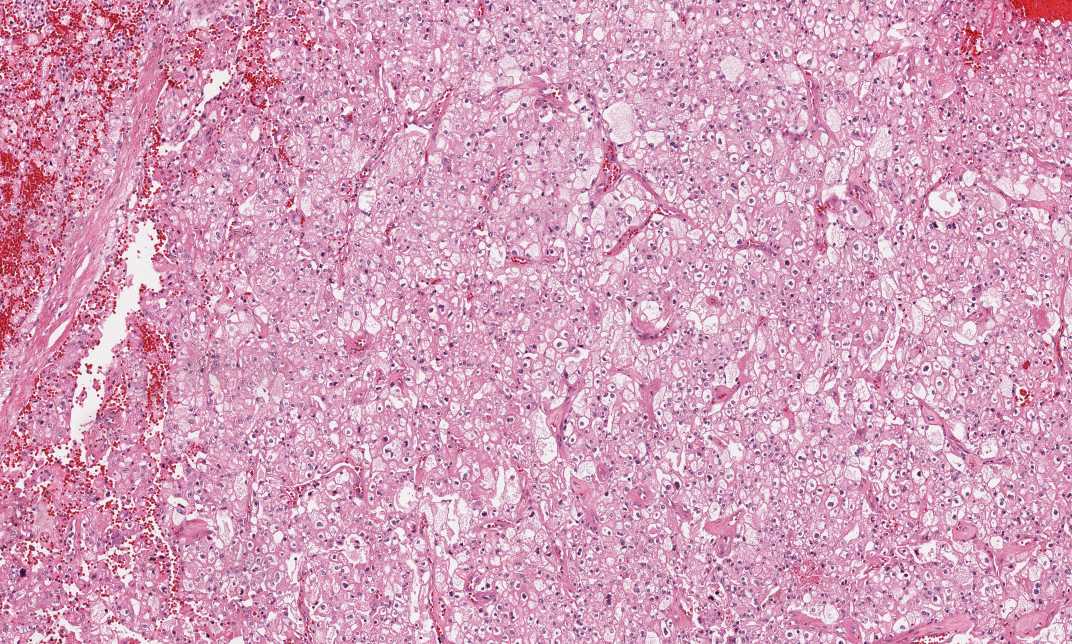
Hematoxylin & eosin |
Area 2: Solid sheets of tumor cells with large polygonal tumor cells, distinct cytoplasmic borders, low-grade nuclei and perinuclear halo. |
|
History: This slide was obtained from an adult. What is this organ and what is your diagnosis?
Histologic Highlights of this Case:
Comment:
|
Bonus Images:
|
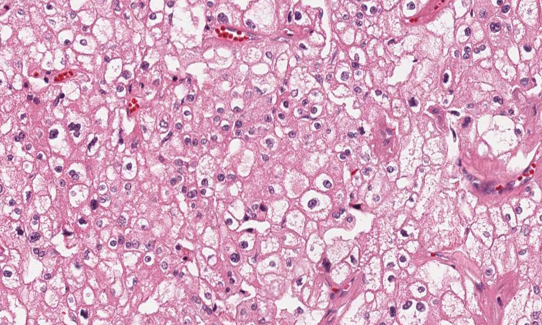
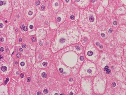
Hematoxylin & eosin |
High magnification: Many of the cells have a fine eosinophilic |
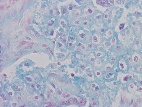 Hale's colloidal Iron |
Colloidal iron: The cytoplasm of this tumor is stained blue with Hale's colloid iron stain. |
Original slide is contributed by Dr. Kar-Ming Fung, University of Oklahoma Health Science Center, Oklahoma, U.S.A.