Case No.: N-024 Quiz
Diagnosis: Glioblastoma, WHO grade IV
Organ: Brain
Last Updated: 3/21/2011
|
|
|
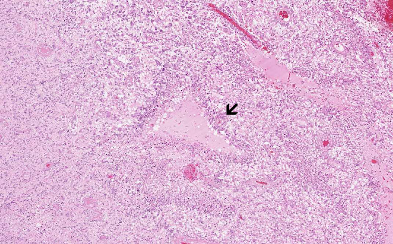
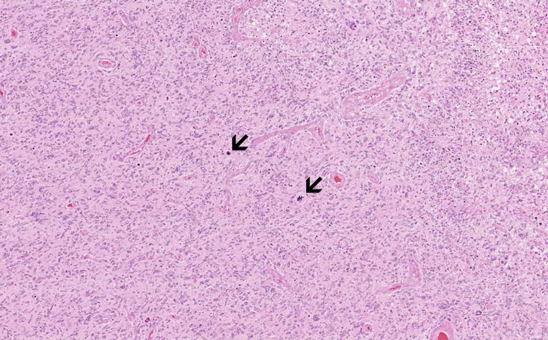
Hematoxylin & eosin |
Area 1: Pseudopalisading necrosis (arrow) is one of the characteristics of glioblasoma. They typically occur as triangular (as illustrated here) or slit like necrosis ( as illustrated in other areas of this slides). Note that there is neoplastic proliferation of glial cells in the background. |
|
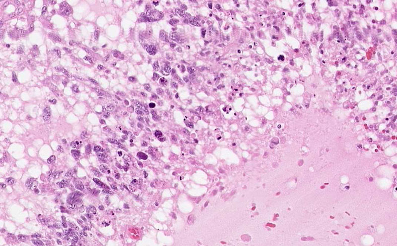
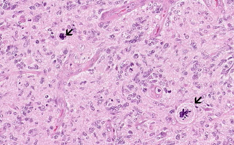
Hematoxylin & eosin |
Area 2: Endothelial proliferation is also a classic feature of glioblastoma (arrow). Note that the vessels has form glomeruloid structures with complex structure and that the entothelial cells are plump and protrude into the lumen of the blood vessels. |
|
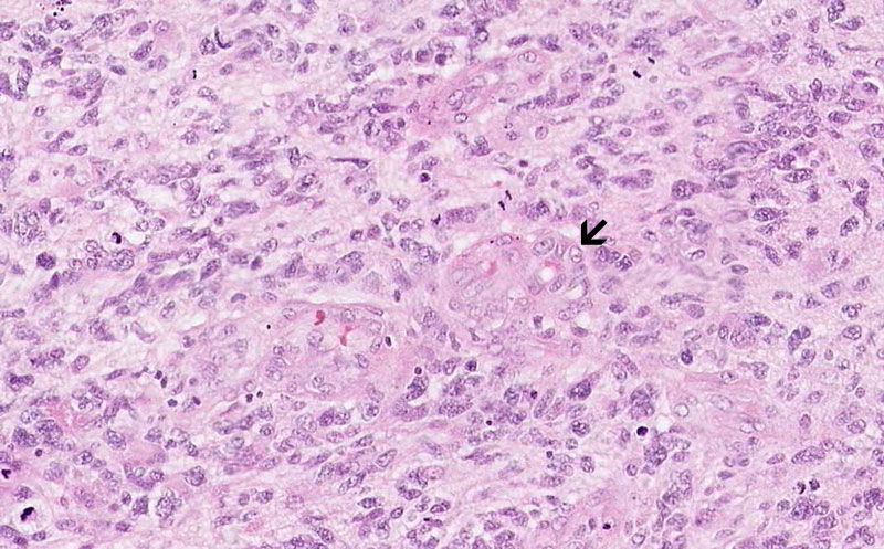
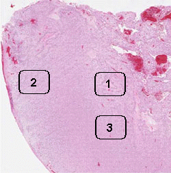
Hematoxylin & eosin |
Area 3: Mitoses, including atypical mitosess (arrow), are common in glioblsastomas. |
|
History: This slide was taken from the archive and the history was not provided. On MRI, this type of lesion typically occur as a ring enhancing lesion with substantial edema and mass effects.
Histologic Highlights of this Case:
|
Original slide is contributed by Fred R Dee MD, Department of Pathology, University of Iowa. (Iowa Image Collection)