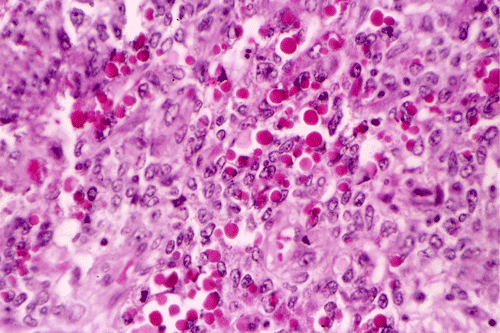
PAS
| An 18 year-old Woman with
a Large Mediastinal Mass. October, 2003, Case 310-3. Home Page |
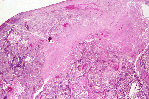
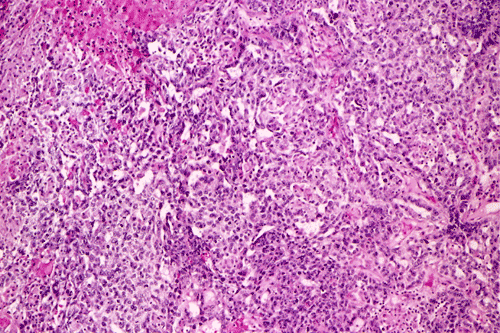
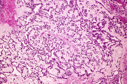
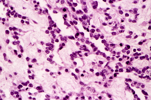
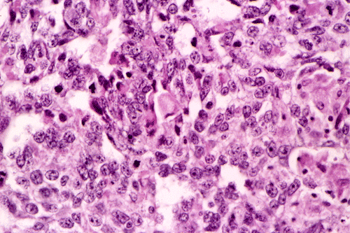
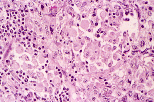
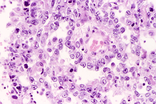
Additional information: A surgery was performed and yielded a 9.5 x 6.5 x 5.5 cm encapsulated mass with attached soft tissue that appeared to be thymic tissue on subsequent microscopic examination. The mass had a gray-yellow, solid cut surface with multifocal hemorrhage and necrosis. The capsule was largely intact with no macroscopic evidence of penetration identified. The following pictures are taken from the surgical specimens.
|
|
|
|
|
|
|
| F. | G. | H. | I. | J. | K. |
|
|
|
||||
| L. |
M. PAS |
PAS: Periodic acid Schiff reaction.
What is your working diagnosis? How would you work this case up further? Back to cytology images Discussion