| A 17 year-old Boy with
Hemoptysis. December, 2003, Case 312-2. Home Page |
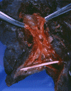
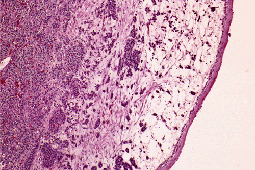
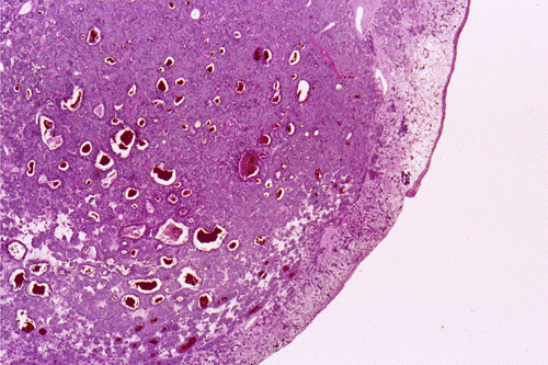
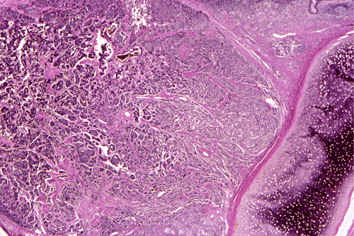
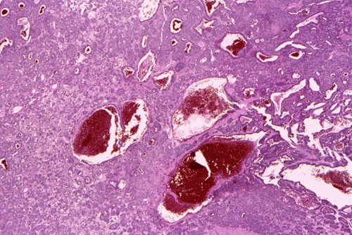
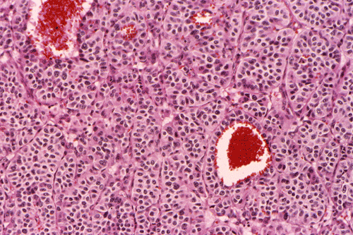
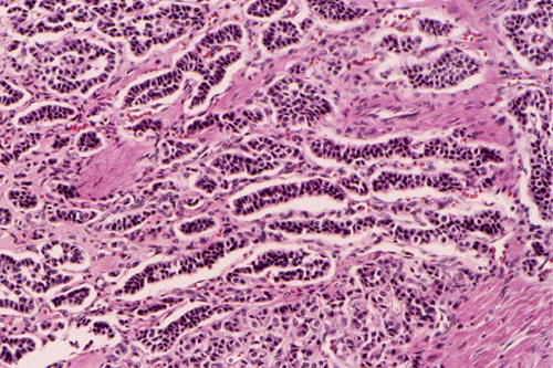
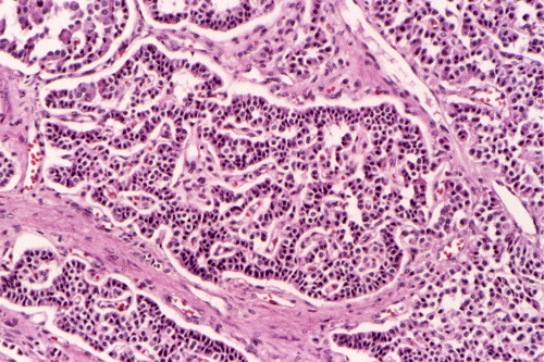
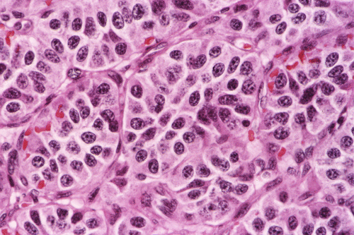
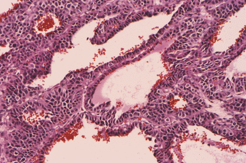
Clinical information: The patient was a 17 year-old boy who was in good health and was an active basket ball player. There was no history of asthma or other major illness. He developed chronic cough, wheezing and shortness of breath about 13 months before presentation. The patient was initially treated in an outside facility under the clinical impression of asthma and his condition improved. Two months before presentation to our institution, he developed fever spikes with worsening of his cough and sputum production. There was also a weight loss of 10 pounds. His pulmonary function tests showed an obstructive pattern with both inspiratory and expiratory abnormal flow volume loops suggestive of variable intrathoracic airway obstruction. The PPD test was negative. A chest x-ray revealed mild atelectasis in the right upper lobe. A bronchoscopy was performed and a biopsy was taken. Based on the pathology results of the biopsy, a lobectomy of the right upper lobe was performed. The following photographs were taken from representative areas of the surgically resected specimen.
|
|
|
|
|
|
|
| A | B | C | D | E | F |
|
|
|
|
|
||
| G | H | I | J |