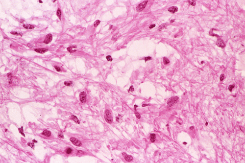
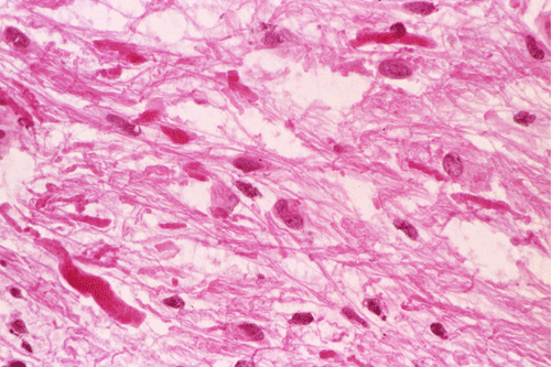
Squash
Squash
Squash
Squash
Frozen
Frozen
Frozen
Frozen
| A 5 year-old Boy with
Nausea and Vomiting for Three Months. January, 2004, Case 401-2. Home Page |
Clinical information:
The patient was a 5 year-old boy who developed intermittent nausea and vomiting. Three days before admission, his conditions deteriorated to the point that he was not able to hold down any food because of regular vomiting. A large mass in the posterior fossa was found on CT scan in an outside hospital. He was transferred to our institution for further evaluation and treatment. On initial admission, the patient was mildly somnolent but could interact normally. He also complained of mild headache. On neurologic examination, he had some difficulties with upward gaze. Otherwise he was neurologically intact. There was no weakness in the extremities.
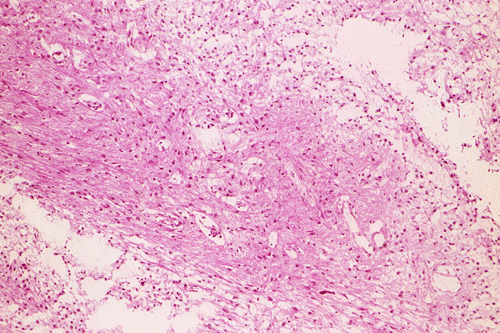
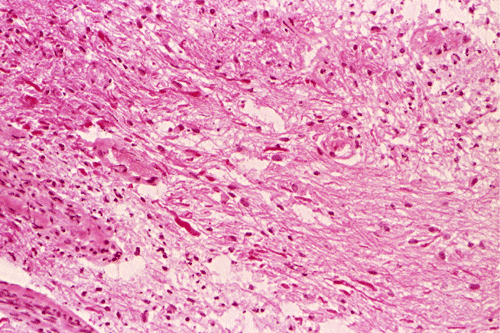
MRI scan was performed and revealed a solitary, large, intraxial mass in the right cerebellar hemisphere. It compressed the 4th ventricle and causing obstructive hydrocephalus of the third and lateral ventricle and there is also mild leftward midline shift. The mass had internal cystic changes and a mural component. The mass was hypointense on T1-, hyperintense on T2 weighed images and was enhancing. Some curvilinear area of decreased signal intensity on both T1- and T2-weighed images suggesting calcifications were present.
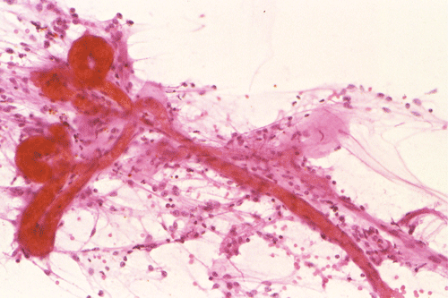
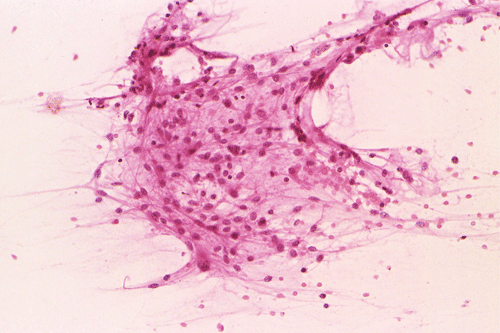
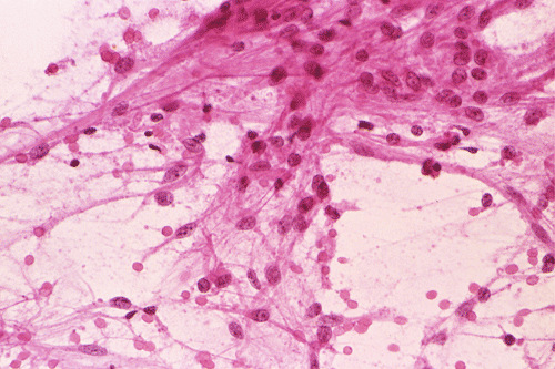
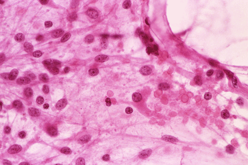
The following pictures were obtained from intraoperative consultation:
|
|
|
|
|
|
|
|
A. Squash |
B. Squash |
C. Squash |
D. Squash |
E. Frozen |
F. Frozen |
|
|
|
||||
|
G. Frozen |
H. Frozen |