
| A 18 month-old Girl with a
Hypothalamic Tumor. March, 2005, Case 503-1. Home Page |
Zhongxin Yu, M.D., M.S.1, Gregory N. Fuller, M.D., Ph.D.2
1 Department of Pathology, University of Oklahoma Health Sciences Center, Oklahoma City, OK and 2 Department of Pathology, University of Texas MD Anderson Cancer Center, Houston, Texas
Clinical information: The patient was an 18 month-old girl who had a contrast enhancing hypothalamic mass. The mass was surgically removed. The following photos are taken from representative areas.
|
|
|
|
|
|
|
| A. | B. | C. | D. | E. | |
|
|
|
|
|
||
| F. | G. | H. | H. |
Pathology of the case:
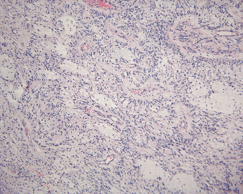
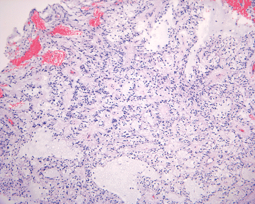
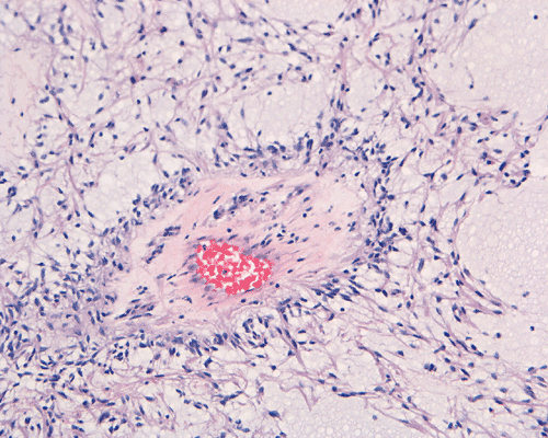
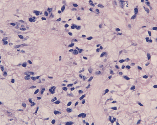
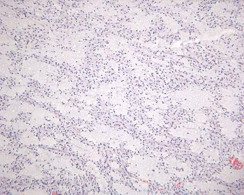
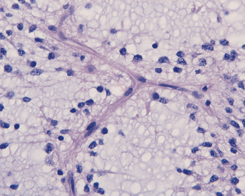
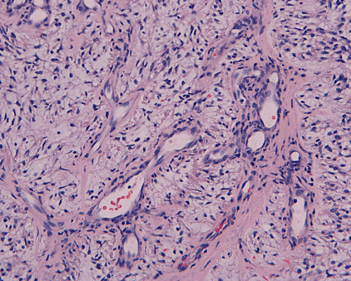
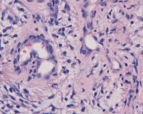
In a substantial amount of area, the tumor is composed of spindle cells with prominent perivascular arrangment and microcyst formation (Panel A and B). These vessels are surrounded by a thin rim of bipolar spindle cells (Panel C). The paucicellular perivascular mantle that is typical for ependymoma is absent in these perivascular arrangements. Adjacent to these perivascular arrangment is substantial amount of myxoid changes. On high-magnification, the tumor cells appear bland in histology, bioplar and spindle in shape, and admixed with a large amount of myxoid substance (Panel D and E). There is a lack of mitosis or significant pleomorphism. In some areas, the tumor is composed exclusively of spindle cells in a myxoid background with microcyst formation but without perivascular coronary arrangement of tumor cells (Panel F). It is not uncommon to observe areas with spindle cells clinging to the blood vessels (Panel G). In a minority of areas, there is increase in cellularity (Panel H). Some vessels seem to be composed of glomeruloids of blood vessels with plump endothelial cells (Panel I).
|
DIAGNOSIS: Pilomyxoid astrocytoma. |
Discussion: General Information Pathology Prognosis Differential diagnosis
General Information
Pilomyxoid astrocytoma (PMA) is a recently described astrocytic neoplasm entity, 1 which has not been included in the current (2000) World Health Organization (WHO) classification 2. This entity typically arises in the hypothalamus or optic chiasm of an infant or young child with the mean age of presentation from 10 to18 months of age (range 2 - 84 month) 1, 3. Occasional PMAs were seen in other locations of the central nervous system including spinal cord or older patients.3, 4, 5.
The most common presenting symptoms of PMA includes failure to thrive, developmental delay, altered consciousness, vomiting, feeding difficulties, and generalized weakness. Radiographically, PMA often occurs as a well-circumscribed, contrast-enhancing mass without substnatial peritumoral edema or parenchymal infiltration. The majority of tumors are solid but a minimal cystic component can be seen in some of these tumors. Some tumor may have focal necrosis or hemorrhage 1, 3, 4, 5, 6, 7. A case of PMA arising in a patient with neruofibromatosis type 1 has been reported in the literature.8 Otherwise, there are no specific cytogenetic, molecular pathologic findings, or specific syndromes that are known to be associated with PMA at this time.
Histologically, the salient features of PMA are rather monotonous, small, spindle bipolar cells with angiocentric arrangement within a strikingly myxoid background. The myxoid basckground material is positive for Alcian blue but negative for PAS 1, 3,. These features are well illustrated in our case. Occasional necrotic foci and mitotic figures can be present. Atypical mitotic figures and substantial nuclear pleomorphism should not be seen. Occasionally, the tumor cells infiltrate the surrounding non-neoplastic brain parenchyma but neither the histological feature nor radiological features would suggest a diffusely infiltrating astrocytoma. In contrast to pilocytic astrocytomas, PMAs do not possess a true alternating densely packed-loosely packed biphasic pattern, do not contain eosinophilic granular bodies or Rosenthal fibers 1, 4. Development of features that are seen in pilocytic astrocytomas such as the biphasic pattern and Rosenthal fibers have been described by Fernandez et al 4. These features are uncommon at the initial presentation but they can develop after chemotherapy. Although these features may suggest maturation of the tumor after chemotherapy, no association with improved prognosis has been described in the study by Fernandez et al 4. The angiocentric arrangement of tumor cells is another trap as it would suggest ependymoma. However, these arrangements are more irregular and fibrillar than the perivascular rosettes in ependymomas.
Immunohistochemically, the tumor cells are strongly positive for glial fibrillary acidic protein (GFAP) and vimentin but they are negative for synaptophysin, neuron specific nuclear protein (Neu-N), chromogranin, epithelial membrane antigen (EMA), and neruofilament. The MIB -1 (Ki-67) labeling index has been reported to vary from 0.9-6.1% with marked regional variations.
Ultrastructurally, the spindle shaped cells contain a large amount of intermediate filaments 8. Fuller et al. described microvilli, cytoplasmic blebs and rare cilia in the bipolar spindle cells and these observations raises the possibility of an ependymal phenotype of PMAs. Cytoplasmic vesicles, coated pits, and occasional synaptoid complexes have also been described 10.
Before PMA was recognized as a separate entity, these tumors were often considered as and treated as a variant of pilocytic astrocytoma. However, PMA has histological features that are quite unique and different from pilocytic astrocytomas. Most importantly, PMA behaves in a more aggressive manner than pilocytic astrocytomas. A local recurrence rate after surgery of 55-76% has been found in the studies byTihan et al.1 , Komotar et al. 3, and Fernendez et al 5. Dissemination through the cerebral spinal fluid (CSF) has been demonstrated in 11-14% the cases. In the study by Tihan et al., 33% of the patients died of disease within 2 years after the diagnosis was rendered. PMA merits recognition as a separate entity and distinguishing PMA from pilocytic astrocytomas is of major prognostic importance.
Differential diagnosis
PMA most typically arises in the hypothalamus or optic chiasm of an infant or young child with a mean age 10 to18 months. It has characteristic histological feature - monomorphous, small, spindle bipolar glial cells in a loose fibrillar and strikingly myxoid background and angiocentric pattern. The characteristic combination of age, location and histological feature limits the main differential diagnoses to pilocytic astrocytoma, ependymoma, and low-grade diffuse astrocytoma.
Pilocytic astrocytoma is an indolent tumor of WHO grade I. The vast majority of them are seen in children. The most common location is the cerebellum but they can be found throughout the brain and spinal cord. Pilocytic astrocytomas arising in optic nerve, optic chiasma/hypothalamus regions are often associated with neurofibromatosis 1. Radiographically, cerebellar pilocytic astrocytomas are typically cystic, with a mural nodule, and enhancing. The salient histologic features are elongated, bipolar cells with long, cytoplasmic process. The tumor cells tend to arrange in a densely-packed, loosely packed pattern. However, other patterns including a clear cell pattern that mimics oligodendroglioma may be present. Rosenthal fibers and eosinophilic granular bodies are often but not always found. Mitotic figures are absent or, at most, uncommon. No substantial nuclear pleomorphism is present. Glomeruloids of thin walled blood vessels are also a common finding. In contrast to PMAs, areas with microcystic changes and myxoid changes are only focal. In the study by Tihan et al., the 13 hypothalamic tumors with classic features of pilocytic astrocytomas shows much better prognosis, no dissemination through the CSF and no patient died of disease 1 . [Click here to see a case of pilocytic astrocytoma]
Ependymoma is a glial neoplasm with phenotypic features of ependymal cells and is a WHO grade II tumor. These tumors occur in both children and adults, Ependymoma can occur at any locations of the brain and spinal cord but are most commonly seen in sites along the ventricular system and in the spinal canal. Histologically, confusion between ependymoma and PMA may occur as both do display perivascular arrangement, bipolar cells, and expression of GFAP. However, the perivascular rosettes of the PMA are more irregular and fibrillar than the perivascular rosettes encountered in ependymoma. True rosettes of cuboidal ependymal cells (ependymal canal) are also seen in ependymoma. The extensive myxoid background is quite unusual for ependymoma. Ependymoma are also positive for epithelial membrane antigen. The specific combination of age, location and histological feature in PMAs is helpful in distinction between these two entities. It is worth to emphasize the importance of distinguishing between these two as the prognosis and treatment for each tumor differ. At present time, PMAs have been mainly treated with surgical resection whereas radiotherapy or chemotherapy is more likely to be considered for ependymoma.
Diffusely infiltrating astrocytomas: diffusely infiltrating astrocytomas is a term applies to a group of astrocytic neoplasms including diffuse astrocytomas (WHO grade II), anaplastic astrocytoma (WHO grade III), and glioblastoma (WHO grade IV). They are usually seen in adults, with diffuse infiltration of adjacent and distant brain structures that is largely irrespective of histological grade. Although myxoid change is quite common in low-grade astrocytoma (WHO grade II), a combination with perivascular arrangement of bipolar cells is not a usual feature of low-grade astrocytomas. In contrast, PMAs are typically present in infant, there is minimal nuclear atypia, and infiltration to the surrounding normal parenchyma, if there is any, are usually focal and minimal. High-grade astrocytomas (anaplastic astrocytomas and glioblastomas) typically show substantial nuclear pleomorphism and other features of malignancy including increased mitotic activity, necrosis, and endothelial proliferation. The high-grade histologic features allows easy distinction from PMAs.
Reference: