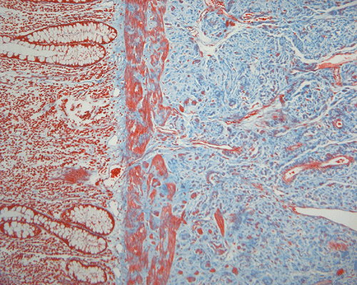
Trichrome
| A 54 year-old Man with a
Colonic Mass and Blood in Stool. February, 2006, Case 6012-2. Home Page |
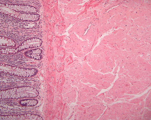
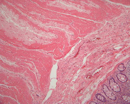
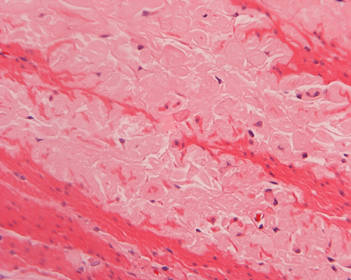
Clinical information: The patient was a 54 year-old who was admitted for evaluation of dysphagia and heartburn with lower gastrointestinal symptoms that included bright-red blood per rectum, mixed with the stool. On physical examination, the patient appeared to be well built and well nourished with no significant findings. On further investigation, he was found to have a mass in the region of the sigmoid colon. Colonoscopy revealed a nearly circumferential large lesion at 30 cm away from the anal verge. The mucosa covering the mass as well as from other part of cecum and colon was otherwise unremarkable. The mass was excised by a sigmoid colectomy.
Grossly, surgical specimen consists of segment of colon, measuring 7.0 cm in length and 6.0 cm in circumference. Mucosal surface reveals a tan, raised, polypoid lesion (4.5 x 3.5 x 2.0 cm), lesion extends into submucosal layer up to 1.5 cm. The followings are representative microphotographs of the sigmoid colon:
|
|
|
|
|
||
| A. | B. | C. |
D. Trichrome |