
Scanne slide
Scanned slide
| A 41 year-old Man with
Headache, Altered Mental Status, and a Large Intraventricular
Hemorrhage. February, 2008, Case 802-1. Home Page |
Clinical information:
The patient was a 41 year-old Hispanic man. One of his friends found him lying in a puddle of blood in a day in May. According to the other information, he had a history of seizure and had been complaining of headache. He started vomiting blood and became unresponsive. He was admitted to the hospital by the EMS.
He lived in a shelter and there was no knowledge of any past medical history, medication, or other details regarding the patient at the time of admission. On physical examination upon admission to the emeregency room, there was dried blood in his nose and his mouth. He was slightly hypertensive and tachycardic. His lung was clear on auscultation and his chest movement was symmetrical. A nasogastric tube was placed and recovered about 200 ml of coffee-ground substance. A CT scan of his head demonstrated a large intraventricular hemorrhage that involved all of the ventricles. The clinical impression was a possible rupture of a distal pericallosal aneurysm. He was sedated and treated for hypertension and possible aspiration pneumonia. Bilateral ventriculostomies were placed in the emergency room.
The patient developed low grade fever in the next several days and his temperature started to spike to around 103 °F about 5 days after admission. Laboratory examination at that time showed elevated WBC, with 64% neutrophils, anemic with Hb 8.4. His CSF cultures were negative for bacteria but his endotrachial aspirate was tested positive for Hafnia alvei (Gram-negative rods). He continued to have tachycardia, tachypnea, hyperthermia, and could not be weaned off from the ventilator. Limited family history was significant for intracranial bleedings in several other family members was subsequently established. His condition continued to deteriorate and he developed metabolic acidosis, hypernatremia, hyperkalemia, elevated ALT and AST, markedly elevated creatine kinase, decreased GFR, acute renal failure, and DIC-like coagulation profile on June 2. Both systolic blood pressure and heart rate dropped to the 60s. Despite all possible measures taken, there was no significant improvement on the patient’s condition. On the 19th hospital day, the patient was made DNR and he expired later that day. An autopsy limited to the brain was performed.
A diagnostic angiography was done during his hospitalization and yielded the following images.
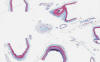
The followings are representative images from autopsy. Panel D to E are taken from the softened area. Panel G and H are taken from the blood vessels of the circle of Willis.
|
|
|
|
|
|
|
| A | B | C | D | E | F |
|
|
|
|
|
||
| G | H |
I Scanne slide |
J Scanned slide |