| A 34 year-old Man with a Cyst in his Left
Maxilla. April, 2020, Case 2004-1. Home Page |
Elizabeth Gillies, M.D., Kar-Ming Fung, M.D., Ph. D.. Last update: April 30, 2020.
Department of Pathology, University of Oklahoma Health Sciences Center, Oklahoma City, Oklahoma.
Clinical information:
The patient was a 34 year-old man. He was referred to our institute because of a cyst in his left maxilla.The patient was otherwise healthy and had no significant family history.
|
|
|
|
||
| A. | B. | C. |
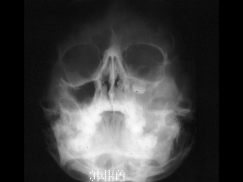
Radiology of the Case:
The sinus on the left side is opacified and contains a tooth. There does not seem to be significant destruction of the surrounding bone on this plain film.
Pathology of the Case:
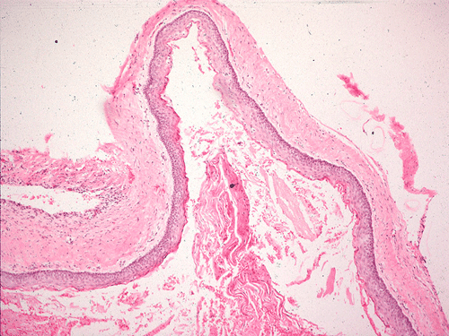
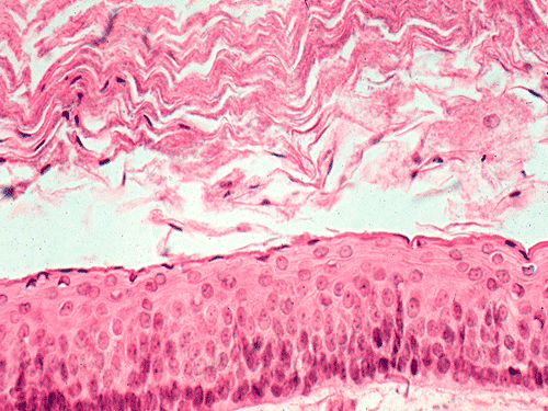
A tooth is present in the specimen. The soft tissue component consists of keratinizing squamous epithelium of rather even thickness of about 5-8 cells thick supported by a fibrous stroma without significant inflammatory cell infiltration (Panel B & C). Keratinaceous debris shredding from the surface of the epithelium is noted. Note that parakeratosis (retention of nuclei in the keratinized layer) is present (Panel C). Good polarization is noted in the epithelium although there are some basal hyperplasia and palisading arrangement of the basal layers. The nuclear looks slightly immature and some with a small but distinct nucleoli. These are features of some low grade dysplasia.
| DIAGNOSIS: Keratocystic odontogenic tumor (odontogenic keratocyst) |
Discussion: General Information Pathology Differential diagnosis Other Cases
General:
Odontogenic keratocyst is better termed keratocystic odontogenic
tumor []
because this entity is locally aggressive and not really a simple cyst.
Recurrence is common. These tumors are mostly seen in the 3rd
and 4th
decades with a 2:1 male predilection. It tends to affect Caucasian subjects.
Posterior mandible is the most common site but any part of mandible and upper
jaw can
be affected. Despite their large size, they are often asymptomatic.
Radiographically, they vary from uniloculated to multiloculated and about
two-thirds of these cases are associated with an impacted tooth. These usually
grow with anterior to posterior extension. Penetration rather than expansion of
the bone is common. Cases may be associated with
nevoid basal cell carcinoma syndrome (Gorlin syndrome)
[Click here to see a case with Gorlin syndrome],
autosomal dominant, associated with PTCH gene and multiple cysts are common in
this situation
[Click here to see a case].
The other findings include bifid rib, calcification of the falx cerebri, ovarian
fibromas, palmar-plantar pitting, and medulloblastoma
[Click here to see a case of medulloblastoma]. Some sporadic cases
without syndromic presentation have loss of heterogosity PTCH on chromosome
9122.3-a31.
Pathology:
This is a cystic neoplasm lined by keratinizing squamous epithelium without atypia, with keratinaceous debris in the lumen similar to that seen in epidermal inclusion cyst. A creamy cystic content may be seen during surgery. The palisading basal layer is a good hint for recognizing this entity. Orthokeratinized odontogenic cysts do not have this palisading arrangement and they have much lower recurrence rate. The parakeratosis also separate this entity from orthokeratinized odontogenic cysts. It can be unilocular to multilocular. It can be inflamed. Marsupialization of the cyst and flushing with caustic agent will change the morphology and lead to a nondescript stratified squamous epithelium with chronic inflammatory cell infiltration in the supporting stroma. Satellite cysts can be seen at the periphery of the tumor and may be related to recurrence therefore these features should be reported. This tumor can perforate bone.
When compared to other odontogenic cysts, the suprabasal cells can be positive for p53 and may have significant amount of Ki-67 labeling. P63 positive cells may extend to one half of the height of the squamous epithelium sparing the parakeratotic epithelial cells. These features may be related to the aggressiveness of this tumor. The superficial squamous cells can be positive for CK4 and CK13. The suprabasal cells can be positive for CK17 and CK19. CD10 can be present [].
Differential Diagnosis:
Dentigerous cysts (follicular cysts) are often found in the same location and
are often associated with an impacted tooth in the same age range. The
epithelium is derived from reduced enamel epithelium and is pluripotent. The
latter can lead to a spectrum of epithelial lining including mucous, ciliated,
and glandular, and salivary gland like cells. Thin, non-keratinizing lining
cells are often seen which can cause confusion with
keratocystic odontogenic
tumor. The epithelial lining however, lacks the palisading basal layer and
keratinaceous content is lacking,
CD10 is typically negative in dentigerous cyst [].
Othokeratinized odontogenic cyst (keratocystic odontogenic tumor) [Click here to see a case] is lined by orthokeratinized squamous epithelium with no palisading of basal layer, no satellite cysts, and lower Ki67 labeling index.
Other Cases: