Horizontal
Sagittal
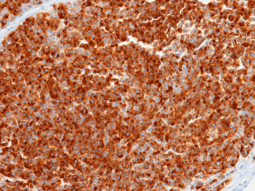
S100
Pan-M
| A 59 year-old Man with a
Maxillary Mass. April, 2007, Case 704-1. Home Page |
Lichao Zhao, M.D., Ph.D., Cheng Z. Liu, M.D., Ph.D., Kar-Ming Fung, M.D., Ph.D. Department of Pathology, University of Oklahoma, Oklahoma City, OK. Last update August 30, 2007.
Clinical information: The patient was a 59 year-old who complained of sinus symptoms including right side nasal congestion for a long but uncertain length of duration. Imaging studies in an outside institute demonstrated a mass in the right maxillary sinus and a biopsy was obtained and the patient was referred to our hospital.
MRI demonstrated a 3.4 cm mass in the right maxillary sinus with heterogeneous enhancement. The mass extend from the inferior anterior to superior anterior aspect of the maxillary sinus with abnormal signal in the anterior wall suggestive of bone invasion. The floor of the orbit was also extensively invaded but no tumor invasion of the orbital content is noted. The cribiform plate does not appear to be involved by tumor. Based on the imaging studies and the biopsy result, a right maxillectomy with orbital exenteration was performed. The following are representative images:
|
|
|
|
|
|
|
|
|
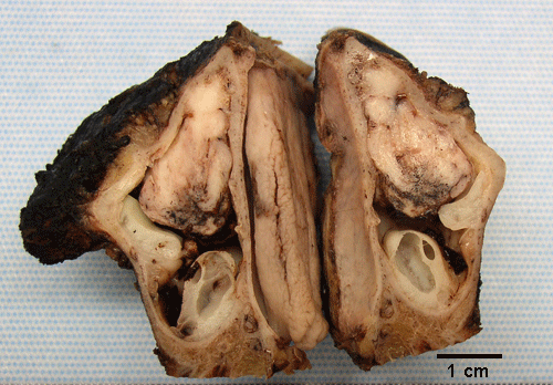
A. Horizontal |
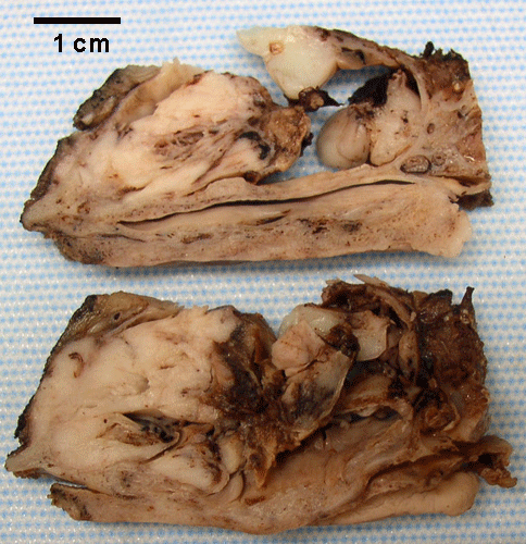
B. Sagittal |
C. | D. | E. | F. | G. |
|
|
|
|||||
|
H. S100 |
I. Pan-M |
Pan-M: HMB45, tyrosinase, and Mart-I.
Histopathology of the case:
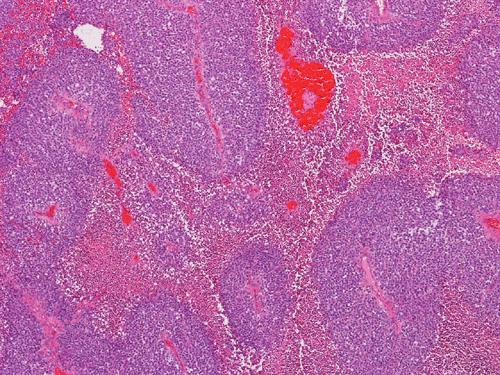
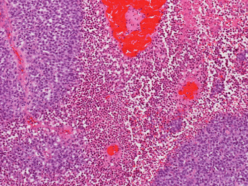
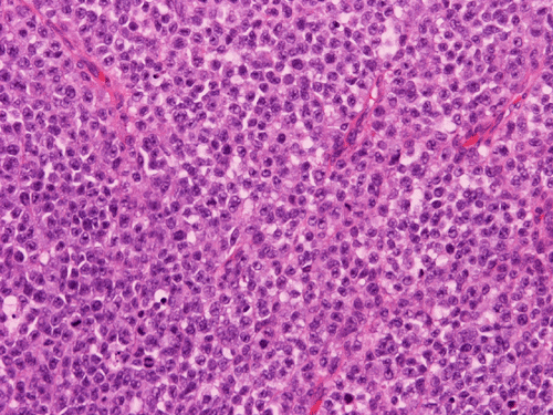
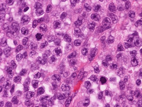
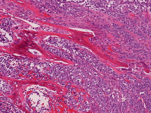
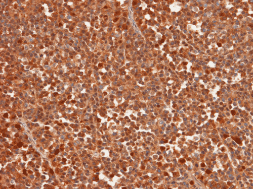
The tumor arises from the anterior half of the maxillary sinus with bone invasion (Panel A and B), Histologically, the tumor is composed of solid sheets of neoplastic cells with extensive necrosis (Panel C and D). On high magnification, the cells appear rather monotonous polygonal cells with indistinct cytoplasmic membrane. The cytoplasm appear slightly bubbly or vesicular with some cells suggestive of clear cells. No genuine clear cells are present. In the viable tumor, brisk mitotic activity are seen. A prominent nuclei is present in many if not all of the nuclei (Panel E and F). Bone invasion is confirmed on histologic sections (Panel G). The tumor cells are strongly positive for S100 and pan-marker (recognize HMB45, tyrosinase, and Mart-1) (Panel H and I).
| DIAGNOSIS: Malignant Melanoma of the maxillary sinus. |
Discussion:
General Information:
The sinonasal tract encompassing the paranasal sinuses (ethmoid, frontal, maxillary, and sphenoid) and the nasal cavities harbor a wide variety of benign and malignant neoplasms. The more common malignant primary tumors tumors in this location include sinonasal undifferentiated carcinoma (SNUC), adenocarcinoma, olfactory neuroblastoma (esthesioneuroblastoma), malignant melanoma, sarcomas, and other rare tumors 1.
Primary sinonasal mucosal melanoma (SNMM) is a rare disease, account for less than 5% of all sinonasal tract neoplasms. They are mostly seen in patients in their 6th or 7th decades. Due to its low incidence, the clinical behavior is not very well studied. The overall prognosis is poor. As per one recent studies of a small series of patients, there is an almost equal predilection between male and female. With treatment, the overall actuarial survival is 49.5% were alive at 2 years and 33.0% at 5 years. Local recurrence and distant metastasis occur early 2. In another study, a median survival of 3 year and a equal predilection between male and female patients are described 3. Formaldehyde exposure and smoking has been suggested as two etiologic factors 4.
Presenting signs and symptoms include nasal obstruction, epistaxis, nasal polyp, pain, nasal discharge, and melanorrhoea. In one study 2, the most common present symptom is epistaxis. At presentation, 70-80% of SNMM cases are localized, 10-20% have regional lymph node and less than 10% have distant metastasis 4. A TNM-type classification separated by anatomic site of involvement and metastatic disease has been proposed to predict biologic behavior 5.
Pathology:
The pathology of malignant melanoma is similar to melanoma arising in other parts of the body. Typically, the tumor is composed of solid sheets of moderately sized to large cells with large nuclei and prominent, eosinophilic nucleoli. Pseudonuclear inclusions are common. The amount of cytoplasm can range from small to moderate and often have a bluish-grayish amphophilic hue. The amount of melanin pigments in cells is extremely variable. It can range from easily found to total absence. The tumor may show solid, papillary, alveolar or sarcomatoid pattern of growth. The tumor cells can be epithelioid, spindled, plasmacytoid, rhabdoid, and/or multinucleated cells. As per one study, SUMM are more likely to have pseudopapillary pattern in comparison to melanoma arising in the oral cavity 3. The cytoplasm may or may not contain melanin pigment. Mitoses are frequent and necrosis is common.
Correct diagnosis of SNMM is not particularly difficult when melanin-rich tumor cells can be identified. However, amelanotic SNMM, particularly those with small cell components can be easily mistaken as other small blue cell tumors. A high index of suspicion is necessary for making the correct diagnosis.
Immunohistochemically, SNMM are positive for S100 and markers for melanomas including HMB45, Mart-1, and tyrosinase. Most of them do not express significant amount of melanin. P16 is expressed in a significant number of these tumors and is mainly related to deletion of 9p21 region 6.
Differential Diagnosis:
The differential diagnosis of SNMM from its mimickers has recently been reviewed by Iezzoni and Mills 7. The followings are some of the more common mimickers.
Malignant lymphoma: Lymphoma presenting in the paranasal sinuses are frequently B-cell lymphomas, with diffuse large B-cell lymphoma (DLBCL) being the most common. The tumor cells are usually dyscohesive and monotonous. The cells usually have round, multicoated or irregularly folded nuclei and multiple small membrane-bound nucleoli or single central prominent nucleolus. DLBCL can be readily distinguished from other tumors by immunohistochemical stains, which show positive reactivity for leukocyte common antigen (LCA) and CD20.
Sinunasal undifferentiated carcinoma (SNUC): The tumor usually grows in nests, lobules, trabeculae and sheets without squamous or glandular differentiation. The tumor cells may show significant pleomorphism and high nuclear-to-cytoplasmic ratios. The nucleoli are variable in size, but most often, they are single and prominent. The mitotic rate is very high and there is often prominent tumor necrosis and apoptosis. This tumor is usually positive for cytokeratin, occasional positive for epithelial membrane antigen, but is usually negative for LCA and S-100.
Olfactory neuroblastoma (esthesioblastoma): This is a malignant neoplasm of neuorectodermal phenotype. In many cases, they occur as small blue cell tumor and some of them may have Homer Wright rosette or Flexner-Wintersteiner rosette. In some tumors, a significant amount of neuropil background can be seen. These tumors are typically positive for synaptophysin. They may also be positive for S100.
Reference:
Stern S. Hanna E. Cancer of the nasal cavity and paranasal sinus. In: Myers E. Suen J. Cancer of the head and neck. Philadelphia: Saunders: 1996:205-233.
Huang SF, Liao CT, Kan CR, Chen IH. Primary mucosal melanoma of the nasal cavity and paranasal sinuses: 12 years of experience. J Otolaryngol. 2007 Apr;36(2):124-9.
Prasad ML, Busam KJ, Patel SG, Hoshaw-Woodard S, Shah JP, Huvos AG. Clinicopathologic differences in malignant melanoma arising in oral squamous and sinonasal respiratory mucosa of the upper aerodigestive tract. Arch Pathol Lab Med. 2003 Aug;127(8):997-1002.
Leon Barnes, John W. Eveson, Peter Reichart, David Sidransky. WHO Classification Head and Neck tumors. lyon, 2005: 72-75
Thompson LD, Wieneke JA, Miettinen M. Sinonasal tract and nasopharyngeal melanomas: a clinicopathologic study of 115 cases with a proposed staging system. Am J Surg Pathol. 2003 May;27(5):594-611.
Franchi A, Alos L, Gale N, Massi D, Paglierani M, Santucci M, Zidar N, Cardesa A. Expression of p16 in sinonasal malignant melanoma. Virchows Arch. 2006 Dec;449(6):667-72.
Iezzoni JC, Mills SE. "Undifferentiated" small round cell tumors of the sinonasal tract: differential diagnosis update. Am J Clin Pathol. 2005 Dec;124 Suppl:S110-21.
Cases of the Month Evaluation Coordinator: KarMing-Fung@ouhsc.edu